Cross-Validation Strategies for Robust Cancer Prediction Models: A Guide for Biomedical Researchers
This article provides a comprehensive guide to cross-validation strategies for developing and validating robust cancer prediction models.
Cross-Validation Strategies for Robust Cancer Prediction Models: A Guide for Biomedical Researchers
Abstract
This article provides a comprehensive guide to cross-validation strategies for developing and validating robust cancer prediction models. Aimed at researchers, scientists, and drug development professionals, it covers foundational principles, methodological applications for various data types (including high-dimensional genomic and clinical data), advanced troubleshooting and optimization techniques, and rigorous comparative validation. The content synthesizes current research to offer actionable insights for mitigating overfitting, assessing model generalizability, and implementing best practices that ensure reliable and clinically translatable predictive models in oncology.
The Critical Role of Cross-Validation in Modern Cancer Prediction
Core Concepts and Definitions
In the development of cancer prediction models, validation is a critical step that ensures the model's findings are reliable and applicable to new patient populations, rather than being artifacts of the specific dataset used for development. Three interconnected concepts are fundamental to this process: overfitting, optimism bias, and generalizability.
Overfitting occurs when a model learns not only the underlying true relationships in the training data but also the random noise specific to that dataset. This is akin to a student memorizing specific exam questions rather than understanding the underlying principles, consequently performing poorly on new questions that test the same concepts. Overfitting is particularly prevalent in high-dimensional settings where the number of potential predictors (e.g., genomic markers) far exceeds the number of observations (patients). This excessive model complexity leads to excellent performance on the training data but poor performance on new, unseen data [1].
Optimism Bias is the direct consequence of overfitting. It refers to the systematic overestimation of a model's predictive performance when evaluated on the same data used for its development. The model's performance appears optimistically good because it has already "seen" this data. The bias is quantified as the difference between the performance on the training data and the expected performance on new, independent data [2]. Mitigating this bias is a primary goal of robust internal validation techniques.
Generalizability (or external validity) describes a model's ability to maintain its predictive accuracy when applied to data from different sources, such as patients from a different geographic region, hospital, or time period. It is the ultimate test of a model's clinical utility. A model that cannot generalize may lead to inaccurate predictions and potentially harmful clinical decisions when implemented in practice [3].
Internal Validation Strategies
Internal validation techniques use the available dataset to estimate and correct for the optimism bias inherent in a newly developed model. The table below summarizes the common strategies, their methodologies, and relative performance based on a simulation study in high-dimensional settings.
| Validation Method | Key Implementation Steps | Stability & Performance Findings (from simulation [4]) |
|---|---|---|
| Train-Test Split | Dataset is randomly split into a single training set (e.g., 70%) and a single test set (e.g., 30%). The model is built on the training set and evaluated on the held-out test set. | Performance was found to be unstable, heavily dependent on a single, arbitrary data split. |
| Bootstrap Validation | Multiple random samples are drawn with replacement from the full dataset to create many bootstrap training sets. Models are built on each and tested on the non-sampled data. | The conventional bootstrap was over-optimistic. The 0.632+ bootstrap variant was overly pessimistic, especially with small sample sizes (n=50 to n=100). |
| K-Fold Cross-Validation | The dataset is partitioned into K equally sized folds (e.g., K=5 or 10). Iteratively, K-1 folds are used for training and the remaining fold is used for validation. This process is repeated K times. | Demonstrated greater stability and is recommended for internal validation of high-dimensional models, particularly with sufficient sample sizes. |
| Nested Cross-Validation | A two-layer procedure. The inner loop performs cross-validation on the training set to tune model parameters (e.g., hyperparameters), while the outer loop provides an almost unbiased performance estimate. | Performance was robust but showed some fluctuations depending on the regularization method used for model development. |
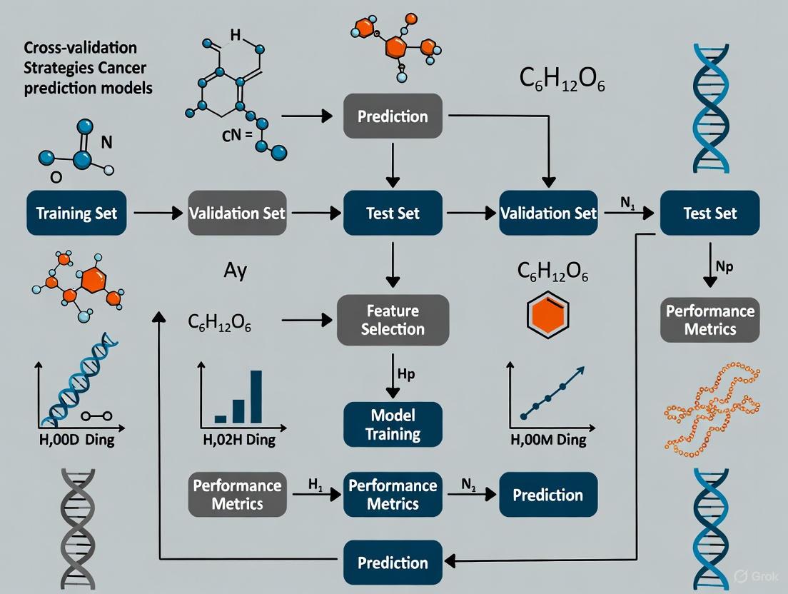
Experimental Protocol for K-Fold Cross-Validation
A commonly used and robust internal validation method is K-Fold Cross-Validation. The following protocol, as applied in a study classifying five cancer types from DNA sequences, details its implementation [5]:
- Data Partitioning: The entire dataset is first divided into a training set and a completely independent hold-out test set (e.g., 80%/20% split). The test set is set aside and not used in any model building or tuning until the final evaluation.
- Stratification: To ensure each fold is representative of the overall class distribution, the training set is partitioned into K folds (typically K=5 or 10) using a stratified sampling approach. This preserves the percentage of samples for each cancer class in every fold.
- Iterative Training and Validation: The following process is repeated K times:
- Training Phase: For each iteration
i(whereiranges from 1 to K), folds 1 through K, excluding foldi, are combined to form a new training subset. - Model Fitting & Tuning: A model is fitted on this training subset. If hyperparameter tuning is required, it is performed within this training subset using a second, inner layer of cross-validation to avoid data leakage.
- Validation Phase: The tuned model is used to predict the outcomes for the data in the held-out fold
i. The performance metrics (e.g., AUC, accuracy) from this prediction are recorded.
- Training Phase: For each iteration
- Performance Aggregation: After K iterations, each data point in the training set has been used exactly once for validation. The K performance estimates are then aggregated (e.g., by averaging) to produce a single, robust estimate of the model's predictive performance, which accounts for optimism bias.
External Validation and Risk of Bias Assessment
While internal validation estimates optimism, external validation is the process of evaluating a model's performance on data that was completely independent of the development process, often collected from different locations or time periods [3]. It is the gold standard for assessing a model's real-world generalizability. For instance, a recent large-scale study developed cancer diagnosis algorithms on a population of 7.46 million patients in England and validated them on two separate cohorts totaling over 5.3 million patients from across the UK, demonstrating superior performance compared to existing models [3].
To systematically evaluate the methodological quality of prediction model studies, the Prediction model Risk Of Bias ASsessment Tool (PROBAST) was developed. This tool is critical for researchers and clinicians to judge the trustworthiness of a published model. PROBAST assesses four domains [6] [7]:
- Participants: Were the participants and the data source appropriate for the research question and representative of the target population?
- Predictors: Were the predictors defined, assessed, and measured in a similar way for all participants?
- Outcome: Was the outcome of interest defined and determined in a robust and consistent manner?
- Analysis: This is the most critical domain. It evaluates issues like sample size, handling of continuous predictors, and most importantly, whether the model was validated appropriately and whether the validation accounted for overfitting and optimism bias.
A systematic review using PROBAST to assess prognostic models in oncology developed with machine learning found that a staggering 84% of developed models were at a high risk of bias, with the "analysis" domain being the largest contributor [7]. Common flaws included insufficient sample size and the use of simple data-splitting without other internal validation techniques, leading to overoptimistic results.
The Scientist's Toolkit: Research Reagent Solutions
The following table details key methodological "reagents" and their functions in the validation of cancer prediction models.
| Research Reagent (Method/Technique) | Primary Function in Validation |
|---|---|
| PROBAST (Prediction model Risk Of Bias ASsessment Tool) | A structured tool to critically appraise a prediction model study for potential methodological shortcomings and risk of bias across participants, predictors, outcome, and analysis domains [6] [7]. |
| Regularization (e.g., Lasso, Ridge) | A statistical technique used during model fitting to reduce model complexity and prevent overfitting by penalizing the magnitude of model coefficients [1]. |
| Bootstrap Resampling | A statistical method that involves repeatedly sampling with replacement from the original dataset. It is used to estimate the distribution of a statistic (e.g., model optimism) and correct for it [4] [2]. |
| Shrinkage | A post-development correction factor applied to model coefficients to make the model's predictions less extreme (more conservative), thereby improving generalizability [2]. |
| Nomogram | A graphical calculating device that provides a visual representation of a multivariate statistical model, enabling clinicians to easily compute an individual patient's predicted probability of an outcome [8]. |
| Grid Search | A hyperparameter optimization technique that systematically works through a manually specified subset of the hyperparameter space to find the combination that yields the best model performance, typically evaluated via cross-validation [5]. |
The Critical Need for Rigorous Validation in High-Dimensional Oncology Data
Modern oncology research increasingly relies on high-dimensional data, where the number of features (such as genomic or transcriptomic variables) vastly exceeds the number of patient samples. While predictive models built from this data hold tremendous promise for personalized cancer care, they are particularly vulnerable to overfitting and optimism bias, where performance estimates on training data are unrealistically high compared to true performance on independent data. This challenge is especially acute with time-to-event endpoints like survival or disease recurrence, where right-censoring adds further complexity [9]. Consequently, rigorous internal validation is not merely a statistical formality but a critical prerequisite for developing reliable models that can genuinely inform clinical decision-making and drug development pipelines.
This guide provides an objective comparison of common internal validation strategies for high-dimensional oncology data, framing them within the broader thesis that cross-validation strategy selection directly impacts performance estimation accuracy and future model utility.
Comparative Analysis of Internal Validation Strategies
A recent simulation study provides a direct benchmark of internal validation methods in a high-dimensional time-to-event setting, typical in oncology. The study simulated datasets inspired by a real-world head and neck cancer cohort, incorporating clinical variables and 15,000 transcriptomic features with realistic distributions [9]. The performance of Cox penalized regression models was assessed using various validation methods, measuring discrimination (time-dependent AUC and C-index) and calibration (3-year integrated Brier Score) across sample sizes from 50 to 1000 [9].
Table 1: Comparison of Internal Validation Method Performance in High-Dimensional Settings
| Validation Method | Key Principle | Stability with Small Samples (n=50-100) | Performance with Larger Samples (n=500-1000) | Risk of Optimism Bias | Recommended Use Case |
|---|---|---|---|---|---|
| Train-Test Split (70:30) | Single split into training and testing sets | Unstable performance | More stable but inefficient data use | Moderate | Preliminary exploration only |
| Conventional Bootstrap | Repeated sampling with replacement | Over-optimistic | Over-optimistic | High | Not recommended |
| 0.632+ Bootstrap | Weighted combination of apparent and bootstrap error | Overly pessimistic | Improves but can remain pessimistic | Low (pessimistic) | Specific scenarios requiring bias correction |
| K-Fold Cross-Validation | Data split into K folds; each fold used once for testing | Good stability | High stability and reliability | Low | Recommended for most scenarios |
| Nested Cross-Validation | Outer loop for performance estimation; inner loop for model selection | Good stability | High stability, but can fluctuate with regularization | Very Low | Recommended when hyperparameter tuning is needed |
The data reveals that k-fold cross-validation and nested cross-validation are the most reliable strategies, offering a superior balance between bias reduction and stability, especially when sample sizes are sufficient [9]. In contrast, simpler methods like train-test splitting or conventional bootstrap resampling demonstrate significant limitations for high-dimensional prognostic models.
Detailed Experimental Protocols and Methodologies
Simulation Framework for Benchmarking
The foundational study for this comparison employed a rigorous simulation protocol to ensure biologically and clinically relevant findings [9]:
- Data Generation Mechanism: Clinical variables (age, sex, HPV status, TNM staging) were simulated based on distributions from the SCANDARE head and neck cohort (NCT03017573). Transcriptomic data for 15,000 transcripts were generated using a four-step process that replicated the mean expression, dispersion, and skewed distribution of real RNA-seq data [9].
- Time-to-Event Simulation: Individual disease-free survival times were generated using an inverted cumulative hazard method. The model incorporated coefficients for clinical variables estimated from the real cohort and assumed 200 of the 15,000 transcripts were truly associated with recurrence risk, with coefficients drawn from uniform distributions [9].
- Experimental Replicates: For each sample size scenario (50, 75, 100, 500, 1000), 100 fully independent dataset replicates were generated and analyzed to ensure robust performance estimates [9].
Model Training and Validation Workflow
The following diagram illustrates the core experimental workflow for training and validating a high-dimensional Cox regression model, as implemented in the benchmark study.
Performance Evaluation Metrics
The benchmark study evaluated model performance using metrics critical for time-to-event data [9]:
- Discrimination: Ability to separate patients with different event times.
- Time-dependent AUC and Harrell's C-index were used. The C-index is a generalization of AUC for censored data.
- Calibration: Agreement between predicted and observed event probabilities.
- Integrated Brier Score (IBS) was used, where a lower score indicates better calibration, especially for 3-year disease-free survival.
Building and validating robust prediction models requires a suite of methodological tools and software resources.
Table 2: Essential Research Toolkit for High-Dimensional Model Validation
| Category | Tool/Reagent | Primary Function | Application Notes |
|---|---|---|---|
| Statistical Methods | Cox Proportional Hazards Model | Models relationship between features and survival time | Foundation for time-to-event analysis [9] [10] |
| Penalized Regression (LASSO, Elastic Net) | Performs variable selection and regularization in high-dimensional settings (p >> n) | Prevents overfitting; improves model sparsity [9] [10] | |
| Validation Algorithms | K-Fold Cross-Validation | Robustly estimates model performance by partitioning data into K subsets | Recommended for stability; balances bias and variance [9] |
| Nested Cross-Validation | Provides unbiased performance estimation when also tuning hyperparameters | Essential for complex model selection [9] | |
| Software & Platforms | R Statistical Software | Open-source environment for statistical computing and graphics | Primary platform used in benchmark study (version 4.4.0) [9] |
| Python with Scikit-Survival | Machine learning library with specialized survival analysis capabilities | Alternative for implementing similar validation workflows | |
| Data Resources | Nationwide Claim Cohorts (e.g., NHIS) | Large-scale, structured data for model development and validation | Enables development of practical, patient-level prediction models [11] |
The empirical evidence clearly demonstrates that the choice of internal validation strategy is not neutral; it fundamentally shapes the perceived and actual performance of high-dimensional oncology prediction models. While k-fold and nested cross-validation currently represent the most reliable approaches, the field continues to evolve. Future research directions include the development of more sophisticated dynamic prediction models that incorporate longitudinal biomarker data to update risk assessments in real-time [10], and the integration of multimodal deep learning frameworks that can effectively combine diverse data types such as clinical, genomic, and imaging data [12]. For researchers and drug developers, prioritizing rigorous validation is a critical investment, ensuring that predictive models translate into genuine clinical utility and advance the frontier of personalized oncology.
In the field of cancer research, the development of robust predictive models using high-dimensional data such as genomics, transcriptomics, and medical imaging has become increasingly prevalent. Internal validation of these models is a critical step to mitigate optimism bias and ensure reliable performance estimates before proceeding to external validation [9]. For researchers, scientists, and drug development professionals, selecting an appropriate validation strategy is paramount, as it directly impacts model generalizability and potential clinical utility. The complex nature of cancer data—often characterized by high dimensionality, limited samples, class imbalance, and correlated features—presents unique challenges that necessitate careful consideration of validation methodologies [13] [9].
This guide provides a comprehensive comparison of common internal validation strategies, with a specific focus on their application in cancer prediction models. We will examine the performance characteristics, implementation requirements, and appropriate use cases for each method, supported by experimental data from recent cancer studies. Understanding these strategies will enable more rigorous model development and more accurate assessment of true predictive performance in oncological applications.
Core Internal Validation Methods
Fundamental Validation Approaches
Internal validation strategies exist on a spectrum from simple holdout methods to sophisticated resampling techniques, each with distinct advantages and limitations in the context of cancer prediction research.
Train-Test Split (also called holdout validation) involves randomly partitioning the available data into separate training and testing sets, typically using a 70-80% portion for model development and the remaining 20-30% for performance evaluation [13] [14]. While computationally efficient and conceptually straightforward, this approach can yield unstable performance estimates, particularly with smaller datasets commonly encountered in cancer studies [9] [15]. For instance, in a mammography radiomics study predicting upstaging of ductal carcinoma in situ, models built from different training sets showed considerable variation, with AUC performances ranging from 0.59-0.70 on training and 0.59-0.73 on test sets across different data splits [15].
K-Fold Cross-Validation addresses some limitations of simple train-test splitting by partitioning the entire dataset into k roughly equal-sized folds (typically k=5 or 10) [14]. The model is trained on k-1 folds and validated on the remaining fold, repeating this process k times with each fold serving as the validation set once [5]. The final performance estimate is calculated as the average across all k iterations. This approach provides more stable performance estimates than single train-test splits and utilizes data more efficiently, making it particularly valuable for smaller cancer datasets [9] [14]. In a study classifying five cancer types using RNA-seq data, 5-fold cross-validation demonstrated excellent stability and achieved a classification accuracy of 99.87% with Support Vector Machines [13].
Stratified K-Fold Cross-Validation is a variant that preserves the class distribution proportions in each fold, which is especially important for cancer datasets with imbalanced outcomes [14]. For example, in a breast cancer classification study, stratified shuffle split cross-validation helped maintain consistent class ratios across splits, contributing to more reliable performance estimation [16].
Nested Cross-Validation employs two levels of cross-validation: an inner loop for hyperparameter tuning and model selection, and an outer loop for performance estimation [9] [4]. This strict separation between model selection and evaluation provides nearly unbiased performance estimates but requires substantial computational resources [14]. In high-dimensional prognosis models for head and neck cancer, nested cross-validation demonstrated good performance, though with some fluctuations depending on the regularization method used for model development [9].
Bootstrap Methods involve repeatedly sampling from the dataset with replacement to create multiple training sets, with the out-of-bag samples used for validation [9]. The standard bootstrap approach tends to be over-optimistic, while the corrected 0.632+ bootstrap method can be overly pessimistic, particularly with small sample sizes (n=50 to n=100) common in cancer studies [9].
Comparative Analysis of Validation Strategies
The table below summarizes the key characteristics, advantages, and limitations of each primary validation method in the context of cancer prediction research:
Table 1: Comparison of Internal Validation Strategies for Cancer Prediction Models
| Validation Method | Key Characteristics | Optimal Use Cases in Cancer Research | Advantages | Limitations |
|---|---|---|---|---|
| Train-Test Split | Single random partition (typically 70/30 or 80/20) | Preliminary model screening with large datasets (>1000 samples) [15] | Computationally efficient; simple implementation | High variance with small datasets; unstable performance estimates [9] [15] |
| K-Fold Cross-Validation | Data divided into k folds; each fold used once as validation | Small to moderate-sized cancer datasets; stable performance estimation [13] [9] | More stable than train-test; efficient data utilization | Can be computationally intensive with large k; requires careful fold creation |
| Stratified K-Fold CV | Preserves class distribution in each fold | Imbalanced cancer outcomes (e.g., rare cancer types) [16] [14] | More reliable for imbalanced data; reduces bias | More complex implementation; requires class labels during fold creation |
| Nested Cross-Validation | Inner loop for model selection; outer for evaluation | High-dimensional settings with hyperparameter tuning [9] [4] | Nearly unbiased performance estimates | Computationally expensive; complex implementation |
| Bootstrap | Multiple samples with replacement; out-of-bag validation | Small datasets where data efficiency is critical [9] | Good statistical properties; confidence intervals | Can be over-optimistic (standard) or pessimistic (0.632+) [9] |
Performance Comparison in Cancer Research Applications
Experimental Evidence from Cancer Studies
Recent research provides compelling experimental data on the performance characteristics of different validation strategies when applied to cancer prediction tasks:
Table 2: Performance Comparison of Validation Methods in Cancer Prediction Studies
| Study Context | Validation Methods Compared | Key Performance Findings | Sample Size | Data Type |
|---|---|---|---|---|
| Head and neck cancer prognosis [9] [4] | Train-test, bootstrap, k-fold CV, nested CV | K-fold and nested CV showed improved stability with larger samples; train-test was unstable; bootstrap was over-optimistic | 50-1000 (simulated) | Transcriptomic (15,000 features) + clinical |
| Breast cancer classification [13] | 70/30 train-test vs. 5-fold cross-validation | SVM achieved 99.87% accuracy with 5-fold CV vs. 96.3% with train-test split | 801 samples | RNA-seq (20,531 genes) |
| Multiple cancer type classification [5] | 10-fold cross-validation with independent test set | 100% accuracy for BRCA1, KIRC, COAD; 98% for LUAD, PRAD with 10-fold CV | 390 patients | DNA sequencing |
| DCIS upstaging prediction [15] | Multiple train-test splits (40 iterations) | AUC varied considerably: training 0.58-0.70, testing 0.59-0.73 across different splits | 700 cases | Mammography radiomics |
| Breast cancer detection [17] | 10-fold cross-validation with multiple splits | Stacked model achieved 100% accuracy using selected optimal feature subsets | 569 patients | Clinical and genomic features |
The experimental evidence consistently demonstrates that cross-validation strategies generally provide more stable and reliable performance estimates compared to single train-test splits, particularly for the high-dimensional, limited-sample datasets common in cancer research [13] [9] [15]. For instance, in a transcriptomic analysis of head and neck tumors, k-fold cross-validation demonstrated greater stability than train-test or bootstrap approaches, especially with larger sample sizes [9]. Similarly, in a breast cancer classification study, models evaluated with 5-fold cross-validation showed approximately 3.5% higher accuracy compared to a simple 70/30 train-test split [13].
Impact of Dataset Characteristics on Validation Performance
The optimal choice of validation strategy depends heavily on dataset characteristics, particularly sample size and dimensionality:
Sample Size Considerations: With smaller sample sizes (n<100), k-fold cross-validation and nested cross-validation generally outperform alternatives, though performance estimates remain variable [9]. As sample size increases to n=500-1000, these methods demonstrate significantly improved stability [9] [15]. In a mammography radiomics study, cross-validation required samples of 500+ cases to yield representative performance estimates [15].
High-Dimensional Data Challenges: Cancer research frequently involves high-dimensional data where the number of features (genes, radiomic features) vastly exceeds the number of samples [13] [9]. In such settings, k-fold and nested cross-validation are recommended as they provide more reliable performance estimates for Cox penalized models [9]. For example, in a study using RNA-seq data with 20,531 genes from 801 samples, 5-fold cross-validation provided stable performance estimates for identifying significant cancer genes [13].
Class Imbalance Issues: Many cancer outcomes exhibit natural imbalance (e.g., rare cancer types, low event rates) [14]. In these scenarios, stratified cross-validation approaches that preserve class distribution across folds are essential to avoid biased performance estimates [16] [14].
Implementation Guidelines for Cancer Research
Methodological Protocols
Based on experimental evidence from recent cancer studies, below are detailed methodological protocols for implementing the most effective validation strategies:
Protocol for K-Fold Cross-Validation in Cancer Transcriptomics [13] [9]:
- Data Preparation: Standardize gene expression data (e.g., RNA-seq counts) using appropriate normalization methods. Check for missing values and outliers.
- Fold Creation: Partition data into k=5 or k=10 folds using stratified sampling based on cancer type or outcome to maintain class distribution.
- Iterative Training/Validation: For each fold iteration:
- Use k-1 folds for feature selection and model training
- Validate on the held-out fold
- Record performance metrics (accuracy, AUC, etc.)
- Performance Aggregation: Calculate mean and standard deviation of performance metrics across all folds.
- Final Model Training: Train the final model on the entire dataset using the optimal hyperparameters identified during cross-validation.
Protocol for Nested Cross-Validation with High-Dimensional Data [9] [4]:
- Outer Loop Setup: Divide data into k outer folds (typically k=5).
- Inner Loop Configuration: For each outer fold, implement an inner cross-validation (e.g., 5-fold) on the training portion.
- Hyperparameter Optimization: Use the inner loop to tune model hyperparameters via grid search or random search.
- Model Evaluation: Train a model with optimized hyperparameters on the inner training set and evaluate on the outer test fold.
- Performance Estimation: Aggregate performance across all outer test folds for an unbiased estimate.
Protocol for Train-Test Validation with Multiple Splits [15]:
- Multiple Iterations: Implement 40-50 random shuffles and splits of the data into training and test sets.
- Balanced Splitting: Maintain consistent outcome rates across training and test splits.
- Performance Distribution Analysis: Record performance metrics for each split and analyze the distribution.
- Stability Assessment: Evaluate the range and variance of performance across splits.
Workflow Visualization
The following diagram illustrates the logical relationship and workflow between different internal validation strategies, highlighting their interconnectedness and appropriate application contexts in cancer research:
This decision framework provides a systematic approach for cancer researchers to select appropriate validation strategies based on their specific dataset characteristics and modeling objectives.
Essential Research Reagent Solutions
The successful implementation of internal validation strategies in cancer prediction research requires specific computational tools and resources. The table below details essential "research reagent solutions" for conducting robust internal validation:
Table 3: Essential Research Reagents for Internal Validation in Cancer Prediction Studies
| Reagent Category | Specific Tools/Libraries | Function in Validation Pipeline | Example Applications in Cancer Research |
|---|---|---|---|
| Programming Environments | Python (scikit-learn, pandas, numpy) [13]; R [9] | Data preprocessing, model implementation, validation execution | RNA-seq analysis [13]; transcriptomic simulation [9] |
| Validation Implementations | scikit-learn crossvalscore, StratifiedKFold [13] [16]; custom nested CV scripts [9] | Automated k-fold, stratified CV, nested CV execution | Breast cancer classification [13] [16]; head and neck cancer prognosis [9] |
| High-Performance Computing | Cloud computing platforms; parallel processing frameworks | Handling computational demands of repeated model fitting | Large-scale transcriptomic analysis [9]; radiomic feature processing [15] |
| Specialized Cancer Datasets | TCGA RNA-seq data [13]; CuMiDa brain cancer expression [13]; MIMIC-III [14] | Benchmark datasets for method development and comparison | Pan-cancer classification [13]; mortality prediction [14] |
| Model Interpretation Tools | SHAP [5]; LIME [17] | Post-validation model explanation and feature importance | DNA sequence classification [5]; breast cancer detection [17] |
These research reagents form the foundation for implementing robust internal validation protocols in cancer prediction studies. The selection of appropriate tools should align with the specific data modalities (genomic, clinical, imaging) and computational requirements of the research project.
Internal validation represents a critical methodological step in developing cancer prediction models that generalize to new patient populations. The experimental evidence and comparative analysis presented in this guide demonstrate that k-fold cross-validation and nested cross-validation generally provide more stable and reliable performance estimates compared to simple train-test splits or bootstrap methods, particularly for the high-dimensional, limited-sample datasets common in cancer research [13] [9].
The choice of optimal validation strategy depends on specific dataset characteristics, including sample size, dimensionality, class balance, and computational resources. For large sample sizes (n>1000), train-test splits may suffice for initial model screening, while small to moderate-sized datasets benefit substantially from k-fold cross-validation [9] [15]. In high-dimensional settings requiring extensive hyperparameter tuning, nested cross-validation provides the most unbiased performance estimates despite increased computational demands [9] [4].
As cancer prediction models continue to evolve in complexity and clinical relevance, employing rigorous internal validation strategies will remain essential for producing trustworthy, generalizable results that can potentially inform clinical decision-making and drug development pipelines.
In computational oncology, the reliable prediction of cancer risk, recurrence, and treatment response is paramount. The performance metrics of these predictive models—often celebrated in research publications—are not inherent properties of the algorithms themselves. Instead, they are profoundly influenced by the choice of validation strategy employed during benchmarking. Benchmarking, the process of evaluating model performance against standardized criteria or datasets to compare different models, serves as the foundation for selecting which models advance toward clinical application [18]. Within this process, the validation strategy—the method for assessing how well a model generalizes to unseen data—acts as a critical filter. It directly controls the reliability of performance metrics such as accuracy, AUC, and hazard ratios. For researchers, scientists, and drug development professionals, understanding this interaction is not merely academic; it is essential for making informed decisions about which models are truly robust enough to trust for preclinical and clinical decision-making.
This guide objectively compares how different validation approaches impact the reported performance of cancer prediction models. It synthesizes findings from empirical benchmarking studies and provides structured protocols to help the research community conduct more rigorous, reproducible, and clinically relevant model evaluations.
Core Principles of Model Evaluation and Benchmarking
Before examining the impact of validation, it is crucial to establish a common understanding of key evaluation concepts and the overarching goals of benchmarking.
Key Model Evaluation Metrics
The performance of predictive models is quantified using metrics that vary based on the task (e.g., classification vs. regression) [19] [20]. The table below summarizes common metrics used in cancer prediction research.
Table 1: Common Evaluation Metrics for Predictive Models
| Metric | Description | Use Case in Cancer Research |
|---|---|---|
| Accuracy | Proportion of total correct predictions [20] | Initial screening of classification models (e.g., cancer type) [5] |
| AUC-ROC | Measures model's ability to separate classes across all thresholds; independent of responder proportion [20] | Overall diagnostic performance (e.g., discriminating cancer vs. normal) [5] |
| Precision | Proportion of positive identifications that were actually correct [19] [20] | When cost of false alarms is high (e.g., recommending an invasive biopsy) |
| Recall/Sensitivity | Proportion of actual positives correctly identified [19] [20] | Critical for screening where missing a case is unacceptable (e.g., early detection) |
| F1-Score | Harmonic mean of precision and recall [20] | Balanced view when class distribution is imbalanced |
| Concordance Index (C-index) | Measures predictive accuracy for time-to-event data (survival analysis) | Assessing recurrence risk models [21] |
| Hazard Ratio (HR) | Ratio of hazard rates between risk groups in survival analysis | Quantifying the separation between high-risk and low-risk patient groups [21] |
The Purpose and Process of Model Benchmarking
Model benchmarking is a structured process for comparing the performance of different machine learning models against a set of standardized criteria or datasets [18]. Its primary purpose is to provide an objective evaluation to determine which model is best suited for a particular task, ensuring that the chosen model meets necessary performance standards before deployment [18]. In cancer research, this is vital for translating algorithms from academic exercises into tools that can genuinely impact patient care.
A robust benchmarking pipeline typically involves several key steps [18]:
- Selection of Benchmark Datasets: Choosing standard, well-characterized datasets that represent real-world inputs.
- Model Training & Evaluation: Training selected models on these datasets and evaluating them using predefined metrics (Table 1).
- Scalability & Efficiency Testing: Assessing computational performance, which is crucial for clinical applications.
- Comparison & Reporting: Systematically comparing results and documenting findings to provide clear recommendations.
Comparing Validation Strategies and Their Impact on Performance
The choice of validation strategy is one of the most consequential decisions in the benchmarking pipeline. Different methods introduce varying levels of bias and variance in performance estimates.
Common Validation Strategies
Table 2: Comparison of Common Model Validation Strategies
| Validation Method | Description | Advantages | Disadvantages & Impact on Performance |
|---|---|---|---|
| Holdout Validation | Dataset is split once into a single training set and a single test set [19]. | Simple and computationally efficient [19]. | High Variance in Metrics: A single, fortunate split can inflate performance. Performance is highly dependent on which samples end up in the test set, leading to unreliable estimates [14]. |
| K-Fold Cross-Validation | Dataset is split into k subsets (folds). The model is trained on k-1 folds and tested on the remaining fold, repeated k times [19] [5]. | More robust performance estimate; uses data more efficiently [19] [14]. | Can be computationally intensive. Subject-wise vs. Record-wise Splitting: In healthcare data, if records from the same patient are split across training and test sets, it can lead to over-optimistic performance due to data leakage [14]. |
| Stratified K-Fold Cross-Validation | A variant of K-Fold that preserves the percentage of samples for each class in every fold [14]. | Essential for imbalanced datasets; provides more reliable estimates for minority classes. | Similar computational cost to standard K-Fold. Mitigates bias in performance metrics that can occur if a random fold contains very few examples of a rare cancer type. |
| Nested Cross-Validation | Features an outer loop for performance estimation and an inner loop for hyperparameter tuning, preventing information leakage between tuning and evaluation [14]. | Considered the gold standard for unbiased performance estimation; reduces optimistic bias [14]. | High Computational Cost. Provides a realistic estimate of how the model will perform on unseen data, often resulting in lower but more trustworthy metrics compared to a single holdout set. |
| External Validation | A model developed on one dataset is tested on a completely independent dataset from a different source or institution [21]. | The strongest test of generalizability; simulates real-world deployment. | Often reveals a significant drop in performance ("performance decay") compared to internal validation, highlighting overfitting to the development dataset's specifics [21]. |
Empirical Evidence: How Validation Choice Affects Metrics in Cancer Research
The theoretical impact of validation strategies is borne out in real-world cancer modeling studies.
Case Study 1: DNA-Based Cancer Classifier. A study developing a high-accuracy DNA-based classifier for five cancer types reported impressive accuracies of up to 100% for some cancer types [5]. However, a closer look at the methodology reveals that these metrics were derived from a 10-fold cross-validation setup on a single cohort of 390 patients [5]. While more robust than a simple holdout, this approach still represents an internal validation. The performance metrics (100% accuracy, AUC of 0.99) are likely optimistic estimates of how this model would perform on DNA data from a different population or sequencing center. Without external validation, the true generalizability of these stellar metrics remains unknown.
Case Study 2: AI for Lung Cancer Recurrence. In stark contrast, a study on an AI model for predicting recurrence in early-stage lung cancer explicitly included external validation [21]. The model was developed on data from the U.S. National Lung Screening Trial (NLST) and then validated on a completely external cohort from the North Estonia Medical Centre (NEMC). The results clearly demonstrate the validation choice's impact: while the model showed strong performance in internal validation (Hazard Ratio for stage I disease: 1.71), its performance was even more pronounced in the external set (HR: 3.34) [21]. This case shows that a rigorous, external validation strategy can not only validate performance but can also strengthen the evidence for a model's utility, providing much greater confidence in its real-world applicability.
The following workflow diagram illustrates how different validation strategies are integrated into a model benchmarking pipeline and how they influence the final performance assessment.
Figure 1: Workflow of validation strategy impact within a benchmarking pipeline. The choice of validation method directly dictates the generated performance metrics and ultimately determines their reliability.
Experimental Protocols for Rigorous Benchmarking
To ensure fair and informative comparisons, benchmarking studies must follow detailed, rigorous experimental protocols.
Protocol 1: Benchmarking with Internal-External Validation
This protocol, inspired by multi-site data studies, provides a robust framework for assessing generalizability when full external validation is not yet possible [14].
- Data Acquisition and Curation: Collect datasets from multiple independent sources (e.g., different hospitals, clinical trials). In a study on a lung cancer AI model, researchers used data from the U.S. National Lung Screening Trial (NLST), North Estonia Medical Centre (NEMC), and the Stanford NSCLC Radiogenomics database [21]. All data must be consistently curated to align clinical metadata and outcomes.
- Site Rotation for Validation: Designate one site's data as the temporary external test set. Pool the remaining sites' data for model training and development.
- Model Development and Tuning: On the pooled training data, perform model training and hyperparameter tuning using an internal method like nested cross-validation to prevent overfitting [14].
- Internal-External Testing: Apply the fully-trained model from Step 3 to the held-out test set from the single site in Step 2. Record all performance metrics.
- Iteration and Meta-Analysis: Repeat Steps 2-4, rotating the held-out test set through each available data site. Finally, aggregate and meta-analyze the performance metrics across all iterations to get a final estimate of out-of-sample performance.
Protocol 2: Large-Scale Benchmarking Tournament
For comprehensively comparing many models, a "tournament" approach, as used in travel demand modeling, can be adapted for cancer informatics [22]. This is suitable for fields with many competing algorithms, such as radiomic feature analysis or genomic biomarker discovery.
- Define the Tournament Arena: Specify the precise prediction task (e.g., recurrence risk within 24 months), the evaluation metrics (e.g., C-index, AUC), and the benchmark datasets.
- Select Competitors: Include a wide range of models, from traditional statistical methods (e.g., Cox regression, logistic regression [23]) to modern machine learning and deep learning models [22]. Ensure all models are evaluated on the same data splits.
- Run Paired Experiments: For each model and dataset combination, run multiple paired experiments using a consistent validation strategy (e.g., repeated k-fold cross-validation) to account for randomness.
- Statistical Comparison: Use a formal statistical model (e.g., a pairwise comparison model) to analyze the results. The goal is to estimate the intrinsic predictive value of each model while controlling for contextual factors like dataset and sample size [22].
- Report and Rank: Report model rankings based on statistical significance, not just point estimates of performance. A key output is to identify a set of top-performing models whose differences are not statistically significant, acknowledging that the "best" model can depend on context.
The Scientist's Toolkit: Essential Reagents for Benchmarking Studies
Table 3: Essential Research Reagent Solutions for Computational Benchmarking
| Tool / Reagent | Function / Purpose | Example Use in Cancer Model Benchmarking |
|---|---|---|
| Standardized Benchmark Datasets | Provides a common ground for fair model comparison. | Publicly available datasets like The Cancer Genome Atlas (TCGA) or MIMIC-III (for critical care) [14] allow different models to be tested on identical data. |
| Stratified K-Fold Cross-Validator | Software function to split data into folds while preserving class distribution. | Prevents optimistic bias from random splits in imbalanced tasks (e.g., predicting a rare cancer subtype) by ensuring all folds have representative examples [14]. |
| Nested Cross-Validation Pipeline | A software script that automates the outer and inner loops of model training and tuning. | Crucial for obtaining unbiased performance estimates when comparing multiple models that require hyperparameter optimization [14]. |
| Radiomics/Feature Extraction Library | Standardized software to quantify medical images into mineable data. | Enables fair comparison of different AI models on the same set of extracted image features (e.g., for predicting lung cancer recurrence from CT scans) [21]. |
| Statistical Comparison Scripts | Code for formal statistical testing of model performance differences (e.g., t-tests, Wilcoxon signed-rank tests). | Moves beyond deterministic claims of "model A beat model B" to statistically sound conclusions about performance superiority in a benchmarking tournament [22]. |
The path to clinically viable cancer prediction models is paved with rigorous benchmarking. As this guide has demonstrated, the reported performance of any model is inextricably linked to the validation strategy used to assess it. A model boasting 100% accuracy under internal cross-validation [5] may see that number plummet upon external validation [21], while a model validated through a rigorous internal-external protocol provides a more trustworthy foundation for further development.
Therefore, the choice of validation is not a mere technicality; it is a fundamental aspect of scientific rigor in computational oncology. By adopting the more demanding practices of nested and external validation, and by embracing comprehensive benchmarking tournaments, the research community can generate more reliable evidence. This will accelerate the translation of truly robust models into tools that can ultimately improve drug discovery and patient outcomes.
In the pursuit of reliable cancer prediction models, researchers consistently face three formidable data challenges: small sample sizes, imbalanced classes, and censored survival data. These issues are not merely statistical nuisances but fundamental obstacles that can skew model performance, generate overly optimistic results, and ultimately limit clinical applicability. Within the broader thesis of cross-validation strategies for cancer prediction research, addressing these data challenges becomes paramount, as the choice of validation methodology is deeply intertwined with data quality and structure. The integration of sophisticated preprocessing techniques with appropriate validation frameworks forms the foundation upon which trustworthy predictive models are built, enabling more accurate stratification of cancer risk, recurrence, and patient survival.
This guide objectively compares contemporary methodologies designed to overcome these data limitations, presenting experimental data and protocols from recent research to inform selection criteria for researchers, scientists, and drug development professionals. By comparing the performance of various techniques on real-world cancer datasets, this analysis provides evidence-based guidance for advancing model robustness in oncological research.
Comparative Analysis of Solutions for Small Sample Sizes
Small sample sizes, particularly prevalent in genomic and rare cancer studies, increase the risk of model overfitting and reduce generalizability. Internal validation strategies become critically important in these high-dimensional, low-sample-size settings.
Internal Validation Strategies for Small Samples
A simulation study based on head and neck tumor transcriptomic data (N=76 patients) provides direct performance comparisons of various internal validation methods in high-dimensional settings. The study evaluated clinical variables and transcriptomic data with disease-free survival endpoints, testing methods across simulated sample sizes from 50 to 1000 patients [4].
Table 1: Performance Comparison of Internal Validation Methods for Small Sample Sizes
| Validation Method | Recommended Sample Size | Stability | Risk of Optimism | Discriminative Performance |
|---|---|---|---|---|
| Train-Test Split | Not recommended for n<500 | Unstable | High | Highly variable |
| Conventional Bootstrap | n=100-500 | Moderate | Overly optimistic | Inflated |
| 0.632+ Bootstrap | n>500 | Moderate | Overly pessimistic | Deflated |
| k-Fold Cross-Validation | n≥50 | High | Well-controlled | Reliable |
| Nested Cross-Validation | n≥75 | High | Well-controlled | Reliable |
Experimental Protocol: k-Fold Cross-Validation for High-Dimensional Data
Research classifying five cancer types (BRCA1, KIRC, COAD, LUAD, PRAD) from DNA sequences of 390 patients demonstrates an effective protocol for small sample sizes using k-fold cross-validation [5]:
- Dataset Partitioning: The entire cohort was divided into training (194 patients), validation (98 patients), and test (98 patients) sets
- Cross-Validation Setup: Implemented 10-fold cross-validation, where the dataset was partitioned into 10 distinct subsets
- Iterative Training: For each iteration, nine subsets (≈194 patients) were used for training, with one subset (98 patients) reserved for validation
- Model Aggregation: Predictions from all 10 validation sets were combined to generate final performance metrics
- Hyperparameter Tuning: Grid search was performed within each fold to optimize parameters without data leakage
This approach achieved remarkable accuracies of 100% for BRCA1, KIRC, and COAD, and 98% for LUAD and PRAD, demonstrating that robust validation can compensate for limited sample sizes [5].
Comparative Analysis of Solutions for Imbalanced Classes
Class imbalance presents a significant challenge in cancer prediction, where minority classes (e.g., malignant cases, rare cancer subtypes) are often the most clinically important. Multiple resampling strategies have been developed to address this issue with varying effectiveness.
Performance Comparison of Imbalance Handling Techniques
Research on colorectal cancer survival prediction using SEER data provides direct comparison of hybrid sampling methods on highly imbalanced datasets (1-year survival imbalance ratio 1:10) [24]. The study evaluated tree-based classifiers with various sampling approaches for 1-, 3-, and 5-year survival prediction.
Table 2: Performance Comparison of Sampling Methods for Imbalanced Colorectal Cancer Data
| Sampling Method | Classifier | 1-Year Sensitivity | 3-Year Sensitivity | 5-Year Sensitivity | Variance Reduction |
|---|---|---|---|---|---|
| None (Baseline) | LGBM | 58.20% | 72.45% | 60.15% | Baseline |
| SMOTE | LGBM | 68.50% | 78.30% | 61.80% | 45.2% |
| RENN | LGBM | 70.10% | 79.95% | 62.40% | 63.7% |
| SMOTE + RENN | LGBM | 72.30% | 80.81% | 63.03% | 88.8% |
| RE-SMOTEBoost | AdaBoost | 75.50%* | 82.30%* | 65.20%* | 88.8% |
Note: *Estimated performance based on reported improvements in original study [25]
Advanced Protocol: RE-SMOTEBoost for Imbalance and Overlap
The novel RE-SMOTEBoost method addresses both class imbalance and overlapping classes through a double-pruning approach [25]:
- Entropy-Based Pruning: Applies information entropy to identify and remove low-information majority class samples across the entire distribution, not just overlapping regions
- Roulette Wheel Selection: Uses Mahalanobis distance and roulette wheel selection to prioritize minority class instances with high information content for synthetic generation
- Boundary-Focused Generation: Generates synthetic samples near decision boundaries using SMOTE, guided by double regularization to prevent new overlapping samples
- Entropy Filtering: Implements a post-generation filter to remove low-quality synthetic samples while retaining informative ones
- Adaptive Boosting Integration: Incorporates the double pruning procedure into AdaBoost, leveraging adaptive reweighting to emphasize hard-to-classify samples
This approach demonstrated a 3.22% improvement in accuracy and 88.8% reduction in variance compared to the best-performing sampling methods [25].
Comparative Analysis of Solutions for Censored Data
Censoring presents unique challenges in cancer survival analysis, where the event of interest (recurrence, death) may not be observed for all patients during the study period. Different statistical approaches address fundamentally different clinical questions.
Analysis Methods for Censored Endpoints
Research on invasive breast cancer-free survival (IBCFS) highlights how different handling methods for second primary non-breast cancers (SPNBCs) – which are excluded from the IBCFS endpoint – address distinct clinical questions and yield different interpretations [26].
Table 3: Comparison of Statistical Approaches for Censored Cancer Endpoints
| Analytical Approach | Clinical Question Addressed | SPNBC Handling | Interpretation | Recommended Use |
|---|---|---|---|---|
| Ignore SPNBCs | Total treatment effect on IBCFS | Events are counted | Estimates overall treatment effect | Primary analysis for most trials |
| Censor SPNBCs | Hypothetical IBCFS risk had no SPNBCs occurred | Patients are censored at SPNBC occurrence | Estimates effect under hypothetical condition | Sensitivity analysis |
| Competing Risks | IBCFS risk while free from any SPNBC | Treated as competing events | Estimates cause-specific effect | When SPNBC risk is high |
External Validation Protocol for Survival Models
A machine learning model for early-stage lung cancer recurrence risk stratification demonstrates rigorous validation methodology for censored data [21]:
- Multi-Cohort Design: Incorporated data from 1,267 patients across U.S. National Lung Screening Trial (NLST), North Estonia Medical Centre (NEMC), and Stanford NSCLC Radiogenomics databases
- Temporal Validation: Used 1,015 patients for algorithm development, with 725 for internal validation and 252 from NEMC as external validation cohort
- Preoperative Focus: Trained survival model using preoperative CT radiomic features and clinical variables to predict recurrence likelihood
- Stratified Performance: Evaluated model using concordance index and disease-free survival across full cohort and within stage I patients specifically
- Pathologic Correlation: Assessed relationship between ML-derived risk scores and established pathologic risk factors using t-tests
The model demonstrated superior performance compared to conventional TNM staging, with hazard ratios of 3.34 versus 1.98 for stratifying stage I patients in external validation [21].
The Scientist's Toolkit: Essential Research Reagents and Solutions
Table 4: Key Research Reagent Solutions for Addressing Cancer Data Challenges
| Reagent/Solution | Primary Function | Application Context | Key Benefit |
|---|---|---|---|
| k-Fold Cross-Validation | Robust performance estimation with limited data | Small sample sizes, high-dimensional data | Minimizes overfitting, maximizes data utility |
| Nested Cross-Validation | Unbiased hyperparameter tuning and validation | Model selection with small samples | Prevents optimistic performance estimates |
| SMOTE + RENN Pipeline | Hybrid resampling for class imbalance | Medical datasets with rare outcomes | Improves sensitivity, reduces variance |
| RE-SMOTEBoost | Advanced ensemble resampling | Combined imbalance and class overlap | Double pruning enhances boundary capture |
| Structural Similarity Score (SSS) | Synthetic data quality assessment | AI-generated synthetic datasets | Validates fidelity to original data distribution |
| Competing Risks Analysis | Accurate time-to-event estimation | Survival data with multiple event types | Prevents biased cause-specific risk estimates |
Based on comparative performance data, researchers can strategically select methodologies based on their specific data challenges:
For small sample sizes (n<100), k-fold cross-validation and nested cross-validation provide the most stable performance, with k-fold being computationally more efficient for initial experiments. When sample sizes exceed 500, the 0.632+ bootstrap method becomes increasingly viable.
For imbalanced classes, the hybrid SMOTE+RENN approach with LightGBM classifiers delivers superior sensitivity for highly imbalanced scenarios (imbalance ratio >1:10), while RE-SMOTEBoost offers additional benefits when class overlap is suspected.
For censored data, the ignore approach for excluded components (like SPNBCs in IBCFS) is recommended for estimating total treatment effects in most clinical trials, with competing risks analysis reserved for high-risk scenarios.
The most robust cancer prediction models will integrate multiple strategies—perhaps combining synthetic data generation for class imbalance with nested cross-validation for small samples—tailored to their specific data limitations and clinical questions. This comparative analysis demonstrates that methodological choices in addressing data challenges significantly impact model performance, reinforcing their critical role within a comprehensive cross-validation strategy for cancer prediction research.
Implementing Cross-Validation for Genomic and Clinical Data
In the field of oncology research, the development of robust and generalizable machine learning models is paramount for accurate cancer prediction and diagnosis. Cross-validation (CV) stands as a critical methodology for reliably estimating model performance, particularly when working with high-dimensional biological data such as genomics, transcriptomics, and histopathological imaging. The core principle of cross-validation involves partitioning a dataset into complementary subsets, performing model training on one subset (training set), and validating the model on the other subset (validation or test set). This process mitigates the risk of overfitting and provides a more realistic assessment of how the model will perform on unseen data. In cancer research, where datasets are often characterized by limited sample sizes alongside a vast number of features (e.g., gene expression data from RNA sequencing), rigorous validation is indispensable for developing trustworthy predictive models [13] [9].
Two predominant cross-validation approaches are K-Fold Cross-Validation and its enhanced variant, Stratified K-Fold Cross-Validation. The fundamental distinction between them lies in how the data is partitioned. Standard KFold divides the data into k consecutive folds after potentially shuffling the data, whereas StratifiedKFold ensures that each fold preserves the percentage of samples for each target class [27] [28]. This preservation of class balance is especially crucial in medical datasets, which frequently exhibit inherent class imbalances, such as a higher proportion of healthy control samples compared to cancer-positive cases. The choice between these two validation strategies can significantly impact performance estimates and, consequently, the perceived success of a cancer prediction model [29] [30].
Theoretical Foundations and Key Differences
K-Fold Cross-Validation
K-Fold Cross-Validation is a foundational resampling technique used to evaluate machine learning models. The procedure is systematic:
- The entire dataset is randomly shuffled (optional but recommended if the data has an inherent order).
- The shuffled dataset is split into k mutually exclusive subsets (folds) of approximately equal size.
- For each of the k iterations, a single fold is retained as the validation data, and the remaining k-1 folds are used as training data.
- The model is trained on the training set and evaluated on the validation set. The performance metric (e.g., accuracy) is recorded.
- After k iterations, the average of the k performance metrics is reported as the overall performance estimate.
A significant characteristic of this method is that each data point appears in the test set exactly once [31]. While KFold is a robust method, its primary drawback emerges with imbalanced datasets: a random partitioning may result in one or more folds having very few or even zero instances of a minority class. This can lead to unreliable performance estimates, as the model cannot be adequately trained or evaluated on underrepresented classes [27] [30].
Stratified K-Fold Cross-Validation
Stratified K-Fold Cross-Validation is an enhancement of the standard KFold method, specifically designed for classification problems. It employs a stratification process, which rearranges the data to ensure that each fold is a good representative of the whole by preserving the original class distribution [28] [30].
For example, consider a binary classification dataset for cancer detection (Class 0: "No Cancer," Class 1: "Cancer") with 100 samples, where 80% are Class 0 and 20% are Class 1. In a 5-fold stratified split, each fold would contain roughly 16 Class 0 samples (80% of the fold size of 20) and 4 Class 1 samples (20% of the fold size of 20). This is in contrast to standard KFold, where a fold might, by chance, contain only 1 or 2 Class 1 samples, or even none at all [30].
This method is widely recommended for classification tasks because it produces more reliable performance estimates, with lower bias and variance compared to regular cross-validation, especially in the presence of class imbalance [28]. Research has demonstrated that stratification is generally a better scheme for accuracy estimation and model selection [28].
Comparative Workflow
The diagram below illustrates the logical sequence and key difference in the splitting mechanism between the two cross-validation strategies.
Figure 1: A comparative workflow of K-Fold and Stratified K-Fold cross-validation, highlighting the key difference in how folds are created.
Experimental Protocols and Performance in Cancer Research
The practical implications of choosing a cross-validation strategy are evident in various cancer prediction studies. Researchers routinely employ these methods to validate models built on diverse data types, from genomic sequences to clinical images.
Experimental Protocol for Model Validation
A standard protocol for implementing these methods in a cancer classification study involves several key steps, as exemplified by research on predicting cervical cancer and classifying multiple cancer types from RNA-seq data [13] [29] [5]:
- Data Preprocessing: This includes handling missing values, outlier removal, and feature scaling. For instance, in a study classifying five cancer types from RNA-seq data, features were scaled using
StandardScalerto normalize the data [5]. - Feature Selection: Given the high dimensionality of omics data, techniques like Lasso (L1 regularization) and Ridge Regression (L2 regularization) are often used to identify the most significant genes or features. Lasso is particularly favored for its ability to drive some coefficients to zero, performing automatic feature selection [13].
- Model Training with Cross-Validation: The dataset is partitioned using either KFold or StratifiedKFold. A common practice is to use a 5-fold or 10-fold setup.
- For a 5-fold CV, the data is split into 5 subsets. The model is trained on 4 subsets (80% of the data) and validated on the remaining 1 subset (20% of the data). This process is repeated 5 times so that each subset serves as the validation set once [13].
- In Stratified K-Fold, this splitting is done while maintaining the original class proportions in each fold [29].
- Performance Aggregation: The performance metrics (e.g., accuracy, precision, recall, F1-score) from each of the k iterations are averaged to produce a single estimate. This average provides a more robust measure of the model's predictive power than a single train-test split [30].
Quantitative Comparison in Research Studies
The table below summarizes the use of cross-validation strategies in recent cancer prediction studies, highlighting their application and resulting performance.
Table 1: Application of Cross-Validation in Recent Cancer Prediction Studies
| Cancer Type / Focus | Data Modality | Validation Strategy | Key Reported Performance | Citation |
|---|---|---|---|---|
| Multiple Cancers (BRCA, KIRC, etc.) | RNA-seq Gene Expression | 5-Fold Cross-Validation | Support Vector Machine achieved 99.87% accuracy. | [13] |
| Cervical Cancer | Clinical Risk Factors | Stratified K-Fold Cross-Validation | Random Forest classifier was identified as a good alternative for early classification. | [29] |
| Cervical Cancer | Diagnostic Images | Stratified K-Fold Cross-Validation | Assisted in evaluating ML models (SVM, RF, etc.) for predicting four common diagnostic tests. | [29] |
| Colon & Lung Cancer | Histopathological Images | 10-Fold Cross-Validation | Used to evaluate a novel LBP method, achieving accuracies up to 96.87%. | [32] |
| Head & Neck Carcinoma | Transcriptomic & Clinical | K-Fold & Nested CV | K-fold CV demonstrated greater stability for internal validation in high-dimensional settings. | [9] |
The consensus from contemporary research is that Stratified K-Fold is the preferred method for classification tasks, including cancer type prediction from genomic or clinical data [13] [29]. Its ability to maintain class distribution across folds prevents scenarios where a fold contains no examples of a rare cancer type, which could lead to overly optimistic or unstable performance estimates. However, for regression problems, such as predicting a continuous outcome like patient survival time, the standard K-Fold approach remains appropriate [28].
Implementing robust cross-validation requires specific computational tools and libraries. The following table details key resources commonly used in cancer prediction research.
Table 2: Essential Research Reagents and Computational Tools for Cross-Validation
| Tool / Solution | Function | Relevance to Cancer Prediction Research | |
|---|---|---|---|
| Scikit-learn (Python) | A comprehensive machine learning library. | Provides the KFold and StratifiedKFold classes for easy implementation of cross-validation, along with numerous algorithms and metrics. |
[27] [30] |
| Lasso (L1) Regression | A feature selection and regularization method. | Identifies the most significant genes from high-dimensional transcriptomic data by shrinking less important coefficients to zero. | [13] |
| StratifiedShuffleSplit | An alternative to StratifiedKFold for repeated random splits. | Useful when a specific test set size is required or for a Monte Carlo-style evaluation, though test sets may overlap. | [31] |
| SHAP (SHapley Additive exPlanations) | An Explainable AI (XAI) technique. | Interprets model predictions by quantifying the contribution of each feature (e.g., a specific gene or clinical variable) to the final output. | [5] [33] |
| R Software / Environment | A programming language for statistical computing. | Widely used for survival analysis and handling high-dimensional omics data, with packages available for various validation methods. | [9] |
The choice between standard and stratified k-fold cross-validation is not merely a technicality but a critical decision that affects the validity of a cancer prediction model. The experimental evidence and theoretical underpinnings strongly support the use of Stratified K-Fold Cross-Validation for all classification tasks, which constitute the majority of cancer prediction problems (e.g., cancer vs. normal, or multi-class cancer typing). It should be the default choice for any imbalanced dataset, ensuring that performance metrics are not skewed by unrepresentative folds.
Conversely, standard K-Fold Cross-Validation remains suitable for regression tasks, such as predicting continuous disease-free survival times, or in scenarios where the dataset is sufficiently large and the target variable is evenly distributed [28]. Furthermore, for data with a temporal component, such as longitudinal patient studies, specialized methods like time-series split are more appropriate than either KFold or StratifiedKFold [31].
In conclusion, within the critical context of cancer research, adopting Stratified K-Fold Cross-Validation is a simple yet powerful step toward developing more reliable, generalizable, and clinically relevant predictive models. It provides researchers with a more trustworthy estimate of how their model will perform in a real-world setting, where dealing with imbalanced class distributions is the norm rather than the exception.
Predictive models using high-dimensional data, such as genomics and transcriptomics, are increasingly used in oncology for time-to-event endpoints like disease-free survival and treatment response [4] [9]. In cancer prediction research, where models developed from molecular data (e.g., 15,000 transcriptomic features) must guide critical clinical decisions, validation strategies become paramount. Internal validation of these models is crucial to mitigate optimism bias prior to external validation, as standard approaches like simple train-test splits can yield overly optimistic performance estimates that fail to generalize to new patient cohorts [4] [9].
The fundamental challenge stems from a methodological flaw: when hyperparameter tuning and performance evaluation are performed on the same data subsets, information "leaks" into the model, creating selection bias and overfitting [34] [35]. This problem is particularly acute in high-dimensional settings where the number of features (p) vastly exceeds the number of samples (n), a common scenario in transcriptomic analysis of tumor samples [4]. Nested cross-validation addresses this vulnerability through a rigorous separation of model selection and model evaluation processes.
Understanding the Nested Cross-Validation Architecture
Nested cross-validation (CV) employs two layers of data partitioning: an inner loop for hyperparameter optimization and model selection, and an outer loop for performance estimation of the selected model. This structure ensures that the test sets used for final evaluation remain completely untouched during the model tuning process, providing an unbiased estimate of how the model will perform on truly independent data [34] [35].
The following diagram illustrates the complete nested cross-validation workflow:
In the architectural flow above, the outer loop systematically partitions the data into training and test folds, while the inner loop further divides each outer training fold to select optimal hyperparameters without ever exposing the outer test fold to the model selection process. This rigorous separation prevents the information leakage that plagues single-layer validation approaches [34] [35].
Comparative Analysis of Internal Validation Strategies for Cancer Models
Quantitative Performance Comparison Across Methods
Recent simulation studies using head and neck cancer transcriptomic data provide empirical evidence for comparing validation strategies. The study simulated datasets with clinical variables (age, sex, HPV status, TNM staging) and transcriptomic data (15,000 transcripts) for disease-free survival prediction, with sample sizes ranging from 50 to 1000 patients [4] [9]. Cox penalized regression was performed for model selection, with multiple validation strategies assessed for discriminative performance (time-dependent AUC and C-index) and calibration (3-year integrated Brier Score).
Table 1: Performance characteristics of internal validation methods in high-dimensional cancer prognosis
| Validation Method | Stability | Optimism Bias | Sample Size Efficiency | Computational Cost |
|---|---|---|---|---|
| Train-Test Split | Unstable, high variance [4] [9] | Moderate to high | Inefficient with limited data | Low |
| Conventional Bootstrap | Moderate | Over-optimistic, particularly with small samples [4] [9] | Moderate | Moderate |
| 0.632+ Bootstrap | Moderate | Overly pessimistic, particularly with small samples (n=50 to n=100) [4] [9] | Moderate | Moderate |
| K-Fold Cross-Validation | High stability with larger samples [4] [9] | Low bias | Efficient across sample sizes | Moderate |
| Nested Cross-Validation | High, though some fluctuations based on regularization [4] [9] | Lowest bias | Requires sufficient samples for reliability | High |
Detailed Methodological Comparison
Train-Test Validation: Simple random splitting (e.g., 70% training, 30% testing) demonstrates unstable performance with high variance across different random splits, making it unreliable for model evaluation in resource-limited settings [4] [9].
Bootstrap Methods: Conventional bootstrap approaches demonstrate significant optimism bias, overestimating model performance, while the 0.632+ bootstrap correction swings to the opposite extreme, becoming overly pessimistic particularly with small sample sizes (n=50 to n=100) common in preliminary cancer studies [4] [9].
Standard K-Fold Cross-Validation: This approach strikes a reasonable balance between bias and variance, showing improved stability with larger sample sizes. However, when used for both hyperparameter tuning and performance estimation, it remains vulnerable to optimism bias as the same data informs both model selection and evaluation [36].
Nested Cross-Validation: By completely separating the hyperparameter optimization phase (inner loop) from the performance estimation phase (outer loop), nested CV provides the least biased estimate of true generalization error, making it particularly valuable for assessing model viability before proceeding to expensive external validation studies [4] [34] [9].
Experimental Evidence: A Simulation Study in Head and Neck Cancer
Methodology and Experimental Protocol
A rigorous simulation study provides concrete evidence for comparing validation strategies in high-dimensional time-to-event settings relevant to cancer prediction [4] [9]. The experimental protocol was designed as follows:
Data Generation: Datasets of varying sample sizes (50, 75, 100, 500, and 1000) were simulated with 100 replicates per scenario, inspired by the SCANDARE head and neck cohort (NCT03017573). Simulated data included clinical variables (age, sex, HPV status, TNM staging) and transcriptomic data (15,000 transcripts) with disease-free survival as the endpoint [9].
Model Development: Cox penalized regression (LASSO, elastic net) was performed for model selection, accounting for the high-dimensional feature space [4] [9].
Validation Strategies Compared: The study compared train-test (70% training), bootstrap (100 iterations), 5-fold cross-validation, and nested cross-validation (5×5) to assess discriminative performance (time-dependent AUC and C-index) and calibration (3-year integrated Brier Score) [4] [9].
Evaluation Metrics: Performance was assessed using discrimination metrics (C-index, time-dependent AUC) that measure the model's ability to separate patients with different outcomes, and calibration metrics (integrated Brier Score) that assess the agreement between predicted and observed event rates [4].
Key Findings and Quantitative Results
The simulation results demonstrated clear differences in validation performance across methods and sample sizes:
Table 2: Simulation results for internal validation methods across sample sizes
| Sample Size | Train-Test | Bootstrap | 0.632+ Bootstrap | K-Fold CV | Nested CV |
|---|---|---|---|---|---|
| n = 50 | Unstable, high variance | Over-optimistic bias | Overly pessimistic bias | Moderate stability | Fluctuations by regularization |
| n = 75 | Unstable, high variance | Over-optimistic bias | Overly pessimistic bias | Improved stability | More consistent |
| n = 100 | Unstable, high variance | Over-optimistic bias | Overly pessimistic bias | Good stability | Reliable estimation |
| n = 500 | Moderate stability | Moderate bias | Moderate bias | High stability | Optimal performance |
| n = 1000 | Moderate stability | Reduced bias | Reduced bias | High stability | Optimal performance |
The results clearly indicate that k-fold cross-validation and nested cross-validation improved performance with larger sample sizes, with k-fold cross-validation demonstrating greater stability across sample sizes. Nested cross-validation showed some performance fluctuations depending on the regularization method but provided the most reliable estimates of generalization error, particularly with sufficient samples (n ≥ 500) [4] [9].
Implementation Framework for Cancer Research Applications
Table 3: Essential research reagents and computational tools for implementing nested CV
| Resource Category | Specific Tools/Functions | Application Context | Implementation Notes |
|---|---|---|---|
| Programming Environments | R (version 4.4.0), Python with scikit-learn | General implementation | R preferred for survival analysis; Python for general ML [4] [34] |
| Core Algorithms | Cox penalized regression (LASSO, elastic net), Random Forest, SVM | High-dimensional time-to-event data, classification | Essential for transcriptomic data with 15,000+ features [4] [9] |
| Hyperparameter Optimization | GridSearchCV, RandomizedSearchCV, Bayesian optimization | Inner loop of nested CV | GridSearchCV most common for comprehensive search [34] [37] |
| Cross-Validation Iterators | KFold, StratifiedKFold, RepeatedKFold | Creating data splits | Stratified variants crucial for imbalanced clinical outcomes [34] [36] |
| Performance Metrics | Time-dependent AUC, C-index, Integrated Brier Score | Time-to-event endpoints | Specialized metrics needed for censored survival data [4] [9] |
| Data Simulation | Custom R scripts based on SCANDARE parameters | Method validation | Enables benchmarking before real data application [4] |
Practical Implementation Protocol
Implementing nested cross-validation requires careful attention to the separation between inner and outer loops. The following Python code illustrates the core implementation using scikit-learn:
For high-dimensional time-to-event data as commonly encountered in cancer research, the implementation would utilize Cox regression models and appropriate survival metrics, typically implemented in R [4] [9].
Based on comprehensive simulation evidence and practical implementation considerations, k-fold cross-validation and nested cross-validation are recommended for internal validation of Cox penalized models in high-dimensional time-to-event settings [4] [9]. These methods offer greater stability and reliability compared to train-test or bootstrap approaches, particularly when sample sizes are sufficient.
Nested cross-validation represents the gold standard for unbiased performance estimation when both model selection and evaluation are required from a single dataset. While computationally intensive, it provides the most realistic assessment of how a model will perform on independent patient cohorts, a critical consideration when developing predictive models for clinical cancer applications.
For research practice, we recommend:
- Using nested cross-validation for final model evaluation when hyperparameter tuning is required
- Employing k-fold cross-validation for routine model development when computational resources are limited
- Avoiding simple train-test splits for high-dimensional cancer data due to instability
- Interpreting bootstrap results with caution, particularly for small sample sizes
This validation rigor ensures that cancer prediction models maintain their performance when deployed in actual clinical validation studies and ultimately for patient care applications.
Within the development of clinical prediction models, particularly in oncology, a critical challenge is ensuring that a model's reported performance reflects its true accuracy when applied to new patients. This discrepancy, known as optimism bias, is especially pronounced in studies with limited sample sizes, a common scenario in cancer research involving novel biomarkers or rare cancer subtypes [38] [39]. Internal validation techniques are therefore essential for obtaining realistic performance estimates.
Among the most effective internal validation methods are bootstrap-based techniques, which leverage resampling to correct for optimism. This guide provides an objective comparison of three prominent bootstrap estimators—Conventional (Harrell's) bias correction, the .632 bootstrap, and the .632+ bootstrap—focusing on their application in small-sample settings typical of cancer prediction model research.
Understanding Bootstrap Validation Methods
The Core Bootstrap Principle
The fundamental idea behind bootstrap validation is to resample the original dataset with replacement to create multiple new datasets of the same size. This process allows researchers to simulate the variation that would be encountered if new samples were drawn from the underlying population. In the context of internal validation, the model development process is applied to each bootstrap sample, and the resulting model is tested on the data not included in that sample (the out-of-bag, or OOB, data) [40]. The average optimism—the difference between performance on the bootstrap sample and the OOB data—is then used to adjust the apparent performance of the model built on the original dataset.
Key Bootstrap Estimators
The three main bootstrap estimators for optimism correction are derived from different conceptual frameworks and weight the apparent and OOB performances differently.
- Conventional Bootstrap (Harrell's Bias Correction): This method provides a direct estimate of optimism by averaging the difference between a model's performance on the bootstrap sample and its performance on the OOB data across many resamples. This average optimism is then subtracted from the model's apparent performance on the original dataset [38]. It is widely adopted due to its straightforward implementation.
- The .632 Bootstrap: This estimator recognizes that the conventional bootstrap might be overly pessimistic because each bootstrap sample contains only approximately 63.2% of the unique observations from the original dataset. It thus creates a weighted average of the over-optimistic apparent performance and the over-pessimistic OOB performance, with weights of 0.368 and 0.632, respectively [38] [40].
- The .632+ Bootstrap: An enhancement of the .632 estimator, the .632+ method addresses a key weakness: its potential for downward bias when the model is severely overfit. It introduces a more complex weighting scheme that accounts for the relative overfitting rate (R), which measures how much worse the OOB performance is compared to the performance of a non-informative model (e.g., an AUC of 0.5). The weight given to the OOB estimate increases as the amount of overfitting increases [38] [40].
The following diagram illustrates the workflow for conducting an internal validation using the bootstrap .632+ estimator, from data resampling to the final performance calculation.
Comparative Experimental Data
Simulation Study Findings
A comprehensive simulation study, which used data from the GUSTO-I trial as a foundation, provides direct comparative data on the three bootstrap methods across various modeling strategies relevant to cancer research, such as logistic regression, stepwise selection, and regularized methods (ridge, lasso, elastic-net) [38].
The table below summarizes the key findings regarding bias and root mean squared error (RMSE) for the C-statistic under different sample size conditions.
Table 1: Comparative Performance of Bootstrap Estimators Under Different Sample Sizes [38]
| Sample Size Scenario | Estimator | Bias Direction | Relative Bias Magnitude | Root Mean Squared Error (RMSE) |
|---|---|---|---|---|
| Large Samples (EPV ≥ 10) | Conventional | Low | Very Low | Low |
| .632 | Low | Very Low | Low | |
| .632+ | Low | Very Low | Low | |
| Small Samples | Conventional | Overestimation | Moderate to High | Moderate |
| .632 | Overestimation | Moderate | Moderate | |
| .632+ | Slight Underestimation | Lowest | Can be higher than others |
Key Conclusions from the Simulation:
- In large-sample settings, all three bootstrap methods are comparable and perform well, effectively correcting for optimism [38].
- Under small-sample conditions, biases become evident. The Conventional and .632 estimators tend to exhibit overestimation bias, particularly as the event fraction in the dataset increases [38].
- The .632+ estimator generally demonstrates the smallest bias in small-sample settings. However, its RMSE can be comparable to or sometimes larger than the other methods, especially when used with regularized estimation methods, indicating a trade-off between low bias and estimation stability [38].
Performance Across Model Building Strategies
The same study also evaluated how the bootstrap estimators perform when combined with different statistical techniques for model development. This is critical for cancer prediction models, where techniques like LASSO are often used for variable selection from a high number of potential predictors.
Table 2: Estimator Performance by Model Building Strategy [38]
| Model Building Strategy | Recommended Bootstrap Estimator | Rationale |
|---|---|---|
| Conventional Logistic Regression | .632+ | Outperforms others in bias reduction for standard models. |
| Stepwise Variable Selection | .632+ | Effective in correcting optimism from the selection process. |
| Firth's Penalized Likelihood | .632+ | Works well with this bias-reducing estimation method. |
| Ridge, Lasso, Elastic-Net | Conventional or .632 | The .632+ estimator can have higher RMSE with these methods. |
Experimental Protocols for Comparison
To ensure the reproducibility of the comparative findings, this section details the core methodologies from the cited simulation studies.
- Data Generation: Simulation data were generated based on the real-world distributions of predictors and outcomes from the Global Utilization of Streptokinase and Tissue plasminogen activator for Occluded coronary arteries (GUSTO-I) trial Western dataset. This ensures the simulation reflects realistic clinical data structures.
- Varied Experimental Conditions: The study was designed to investigate the impact of key factors:
- Events per variable (EPV): Ranged from low (e.g., 5) to high (≥10).
- Event fraction: Varied to represent different outcome prevalences.
- Number of candidate predictors.
- Magnitude of regression coefficients.
- Modeling Strategies Tested: For each condition, models were built using:
- Conventional logistic regression (maximum likelihood).
- Stepwise variable selection (using AIC).
- Firth's penalized likelihood method.
- Ridge, Lasso, and Elastic-Net regression (with tuning parameters determined via 10-fold cross-validation).
- Validation Cycle: For each generated dataset and modeling strategy, the apparent C-statistic was calculated, followed by optimism-corrected C-statistics using the Conventional, .632, and .632+ bootstrap estimators. This process was repeated across multiple simulation runs to compute average bias and RMSE.
- Classifier and Metric: The study focused on predicting the performance of a neural network classifier using the Area Under the ROC Curve (AUC).
- Resampling Methods Compared: The .632 and .632+ bootstrap methods were evaluated against leave-one-out cross-validation and other bootstrap variants.
- Key Finding: Under conditions of high feature space dimensionality and small sample size, a large difference in the accuracy of performance prediction was observed between different resampling methods. The .632 and .632+ bootstrap methods were found to perform better than other methods in terms of Root Mean Squared Error (RMSE) for many of the studied conditions [41].
The Scientist's Toolkit
Implementing these bootstrap methods requires specific computational tools and resources. The following table details key "research reagents" for conducting such an analysis.
Table 3: Essential Reagents and Computational Tools for Bootstrap Validation
| Item Name | Function / Description | Example Use in Protocol |
|---|---|---|
| R Statistical Software | An open-source programming language and environment for statistical computing and graphics. | The primary platform for implementing data simulation, model fitting, and bootstrap validation [38]. |
rms Package (R) |
A comprehensive package for regression modeling, strategies, and validation. | Used to implement Harrell's conventional bootstrap bias correction [38]. |
glmnet Package (R) |
A package for fitting regularized linear models via penalized maximum likelihood. | Used to implement Ridge, Lasso, and Elastic-Net regression models within the bootstrap loops [38]. |
| Simulated Datasets | Data generated from known parameters and real-world data structures (e.g., GUSTO-I). | Serves as a gold standard for evaluating the true performance and bias of validation estimators [38] [39]. |
| High-Performance Computing Cluster | A set of computers linked together to handle computationally intensive tasks. | Essential for running extensive simulation studies and a large number of bootstrap replicates (e.g., 100-1000+) in a feasible time [38]. |
The choice of a bootstrap estimator for validating cancer prediction models is not one-size-fits-all and should be guided by sample size and the chosen modeling strategy.
- For large-scale studies (EPV ≥ 10), any of the three bootstrap methods provides a reliable and largely unbiased correction for optimism. The Conventional bootstrap is a reasonable choice for its simplicity.
- For small-sample studies, which are prevalent in cancer research, the .632+ estimator is generally recommended due to its superior bias correction, especially when using conventional logistic regression or stepwise selection [38].
- An important caveat is that when employing regularized regression methods (like Lasso or Ridge), the .632+ estimator's advantage may diminish due to its potentially higher variance. In such cases, the Conventional or standard .632 estimator might be preferred for a more stable estimate [38].
Ultimately, while internal validation via bootstrapping is a powerful and necessary step, it does not replace the need for external validation on fully independent datasets to ensure a model's generalizability to new patient populations.
Predictive models using high-dimensional transcriptomic data are increasingly used in oncology for time-to-event endpoints, such as disease-free survival in cancer patients [42]. Internal validation of these models is crucial to mitigate optimism bias prior to external validation, a common challenge in high-dimensional settings where the number of features (p) far exceeds the number of observations (n) [43] [44]. Cross-validation (CV) strategies provide a robust framework for performance estimation, hyperparameter tuning, and model selection, helping to prevent overfitting and generate reliable, generalizable predictors for clinical applications [45].
This case study focuses on the application of k-fold and nested cross-validation within high-dimensional survival analysis, using a simulation study based on transcriptomic data from head and neck tumors as a representative example [42]. We compare these methods against alternative validation strategies and provide a detailed examination of experimental protocols, performance outcomes, and practical implementation guidelines for researchers in cancer biomarker discovery.
Key Validation Strategies
In high-dimensional survival analysis, several resampling methods are employed to estimate model performance and optimize parameters. The most common strategies include:
- Train-Test Split: The dataset is randomly divided into a single training set (e.g., 70%) and a hold-out test set (e.g., 30%) [42].
- Bootstrap Methods: Multiple samples are drawn with replacement from the original dataset to create training sets, with the out-of-bag samples used for testing. Variants include the conventional bootstrap and the 0.632+ bootstrap [42].
- k-Fold Cross-Validation: The dataset is partitioned into k disjoint folds (typically k=5 or k=10). Iteratively, k-1 folds are used for training and the remaining fold for testing [42] [45].
- Nested Cross-Validation: A two-level procedure featuring an outer loop for performance estimation and an inner loop for hyperparameter tuning and model selection on the training folds [46].
Comparative Performance in High-Dimensional Settings
A simulation study by Dubray-Vautrin et al. (2025) provides a direct comparison of these methods in a transcriptomic survival context, using data from the SCANDARE head and neck cohort (n=76 patients) [42]. The study simulated datasets with clinical variables, transcriptomic data (15,000 transcripts), and disease-free survival, assessing discriminative performance via time-dependent AUC and C-index, and calibration via the 3-year integrated Brier Score.
Table 1: Performance Comparison of Internal Validation Strategies in High-Dimensional Survival Analysis [42]
| Validation Method | Sample Size (n=50-100) | Sample Size (n=500-1000) | Stability | Remarks |
|---|---|---|---|---|
| Train-Test Split | Unstable performance | Performance stabilizes | Low | Highly dependent on a single data split; not recommended for small samples. |
| Conventional Bootstrap | Over-optimistic | Less biased | Medium | Tendency for excessive optimism, especially with small samples. |
| 0.632+ Bootstrap | Overly pessimistic | More realistic | Medium | Corrects for optimism but can be too pessimistic with small n. |
| k-Fold Cross-Validation | Good performance | Improved performance | High | Recommended; demonstrates greater stability. |
| Nested Cross-Validation | Good performance | Improved performance | Medium-High | Recommended; performance can fluctuate with the regularization method. |
Experimental Protocol: A Reproducible Workflow
The following workflow and experimental protocol are synthesized from the cited case study and related literature on best practices [42] [44].
Experimental Workflow
The diagram below outlines the key stages of a robust analytical pipeline for high-dimensional survival prediction.
Detailed Methodological Steps
Data Preprocessing and Normalization
Proper normalization is critical for transcriptomic data. The recommended approach involves:
- Conversion to TPM/FPKM: Transform raw counts to Transcripts Per Million (TPM) or Fragments Per Kilobase Million (FPKM) to correct for technical biases like library size and RNA composition [47] [48].
- Centered Log-Ratio (CLR) Transformation: Apply CLR to address the compositional nature of RNA-seq data. This transformation converts multiplicative relationships between relative abundances into linear relationships, making data more suitable for linear statistical models [48].
- Filtering and Batch Correction: Remove lowly expressed genes and adjust for technical variation introduced by experimental batches using methods like ComBat or surrogate variable analysis (SVA) [47].
Model Training and Feature Selection
Cox penalized regression models, such as Lasso (L1), Ridge (L2), or Elastic Net (combined L1 and L2 regularization), are standard for high-dimensional survival data [42] [49] [44]. These methods perform variable selection and regularization simultaneously to prevent overfitting.
Hyperparameter Optimization
In nested CV, an inner loop is dedicated to optimizing hyperparameters (e.g., the regularization strength λ in penalized models). This is typically done using a separate k-fold CV on the training fold of the outer loop, ensuring the test data in the outer loop remains completely unseen during this process [46] [44].
Performance Evaluation
Model performance is assessed using metrics appropriate for survival analysis:
- Discriminative Performance: Measured by the C-index (Concordance index) or time-dependent AUC, which evaluate the model's ability to correctly rank survival times [42] [49] [44].
- Calibration Performance: Assessed using the Integrated Brier Score, which measures the average squared difference between observed survival status and predicted survival probabilities [42].
The Nested vs. Standard k-Fold Cross-Validation
Conceptual Framework
The fundamental difference between standard and nested cross-validation lies in their structure and purpose, particularly in handling hyperparameter tuning.
Comparative Advantages and Applications
Table 2: Comparison between Standard k-Fold and Nested Cross-Validation
| Aspect | Standard k-Fold CV | Nested k×k-Fold CV |
|---|---|---|
| Primary Purpose | Performance estimation of a model with fixed hyperparameters. | Unbiased performance estimation when hyperparameters need to be tuned. |
| Structure | Single loop: data split into k folds; each fold serves as a test set once. | Two loops: outer loop for performance, inner loop (on training fold) for tuning. |
| Risk of Bias | High if hyperparameters are tuned on the entire dataset before CV. | Low, as the test set in the outer loop is never used for tuning. |
| Computational Cost | Lower. | Significantly higher (e.g., 5x5-fold NCV requires 25 model fits). |
| Recommended Use Case | Final model evaluation after hyperparameters have been fixed. | Algorithm selection and for obtaining a nearly unbiased performance estimate. |
The Scientist's Toolkit: Essential Research Reagents and Computational Solutions
Implementing a robust survival analysis pipeline requires both computational tools and methodological rigor. The following table details key components.
Table 3: Essential Tools and Resources for Transcriptomic Survival Analysis
| Tool / Resource | Type | Primary Function | Remarks / Application in CV |
|---|---|---|---|
| R Statistical Software | Programming Environment | Data preprocessing, statistical analysis, and visualization. | The primary environment for many biostatistical analyses. |
| Python (scikit-survival) | Programming Library | Machine learning and survival analysis. | Offers implementations of CV and penalized Cox models. |
| BRB-ArrayTools | Software Package | Statistical analysis of genomic data. | Includes specialized tools for cross-validated survival risk classification [43]. |
| SurvRank R Package | Software Package | Feature selection for high-dimensional survival data. | Implements a repeated nested CV framework for unbiased feature ranking [44]. |
| NACHOS/DACHOS | Computational Framework | DL model evaluation with NCV and HPC. | Integrates nested CV with automated hyperparameter optimization on high-performance computers [46]. |
| Penalized Cox Models | Statistical Method | Regularized regression for survival data. | The core modeling technique (e.g., Cox Lasso, Ridge, Elastic Net) [42] [49]. |
| C-Index / AUC | Performance Metric | Evaluates model discrimination. | The key metric for assessing predictive performance in CV [42] [44]. |
This case study demonstrates that k-fold and nested cross-validation are recommended internal validation strategies for Cox penalized models in high-dimensional time-to-event settings, offering greater stability and reliability compared to train-test or bootstrap approaches [42]. The simulation study on head and neck tumor transcriptomics shows these methods effectively mitigate optimism bias, a critical requirement for developing trustworthy predictive biomarkers in oncology.
For researchers, the choice between standard and nested CV should be guided by the study's goal: use standard k-fold CV for evaluating a finalized model with fixed parameters, and nested CV for the combined process of algorithm selection, hyperparameter tuning, and obtaining a realistic performance estimate for the entire modeling process [46] [44]. As the field progresses towards more complex models and the integration of multi-omics data, these rigorous validation frameworks will be indispensable for translating computational predictions into clinically actionable tools.
The application of machine learning (ML) to genomic data presents unique challenges, primarily due to the high-dimensionality of features and the frequent class imbalance in medical datasets. Stratified k-fold cross-validation has emerged as a critical methodology to ensure reliable performance estimation under these conditions. This case study examines its pivotal role in developing a DNA-based multi-cancer classifier, demonstrating how this validation strategy ensures model robustness and generalizability for clinical application.
Experimental Protocols & Methodologies
Dataset Composition and Preprocessing
The foundational study for this case study classified five distinct cancer types—BRCA1 (Breast Cancer gene 1), KIRC (Kidney Renal Clear Cell Carcinoma), COAD (Colorectal Adenocarcinoma), LUAD (Lung Adenocarcinoma), and PRAD (Prostate Adenocarcinoma)—using DNA sequences from 390 patients [5].
Key Preprocessing Steps: The raw data underwent several critical preprocessing steps before model training [5]:
- Outlier Removal: Executed using the Pandas
drop()function to eliminate rows containing outliers. - Data Standardization: Performed using
StandardScalerwithin the Python programming environment to normalize feature scales. - Feature Retention: Notably, feature extraction was not conducted; all available features within the dataset were retained and utilized without reduction.
Implementation of Stratified k-Fold Cross-Validation
The research employed a 10-fold cross-validation technique to rigorously evaluate model performance [5]. The core principle of stratified k-fold is to preserve the original class distribution in each subset, which is vital for imbalanced medical datasets [29].
Specific Workflow [5]:
- The entire dataset was partitioned into 10 distinct subsets.
- During each of the 10 training iterations, nine subsets (collectively containing data from 194 patients) were used for training.
- The remaining single subset (comprising 98 patients) was used for validation.
- This cycle was repeated 10 times, with each subset serving as the validation set exactly once.
- Model selection and hyperparameter tuning were performed using stratified 10-fold cross-validation on the training set, with a strict protocol to prevent data leakage between training and validation splits.
- For the final assessment of generalization performance, an independent hold-out test set (20% of the full cohort) was set aside before any model fitting or parameter tuning.
Machine Learning Models and Hyperparameter Optimization
The study developed and compared a suite of machine learning models [5]:
- Individual Algorithms: Logistic Regression, Gradient Boosting, and Gaussian Naive Bayes.
- Blended Ensemble: A novel approach that combined Logistic Regression with Gaussian Naive Bayes.
- Hyperparameter Tuning: A grid search technique was utilized for hyperparameter fine-tuning to optimize model performance.
Comparative Performance Analysis
Quantitative Results of the DNA-Based Classifier
The blended ensemble model, optimized via grid search and validated through stratified k-fold, demonstrated exceptional performance in classifying the five cancer types [5].
Table 1: Classification Accuracy by Cancer Type
| Cancer Type | Abbreviation | Classification Accuracy |
|---|---|---|
| Breast Cancer gene 1 | BRCA1 | 100% |
| Kidney Renal Clear Cell Carcinoma | KIRC | 100% |
| Colorectal Adenocarcinoma | COAD | 100% |
| Lung Adenocarcinoma | LUAD | 98% |
| Prostate Adenocarcinoma | PRAD | 98% |
The model achieved a micro- and macro-average ROC AUC of 0.99, indicating superb overall discriminative ability. The blended ensemble was shown to outperform each individual algorithm and surpass existing state-of-the-art methods, providing a "lightweight, interpretable, and highly effective tool for early cancer prediction" [5].
Comparison with Alternative Modeling Approaches
Other studies utilizing DNA methylation data for cancer classification provide a useful context for comparing different ML architectures and their performance.
Table 2: Performance of Alternative Classifiers on DNA Methylation Data
| Study Focus | Model | Key Performance Metric | Stratified k-Fold Application |
|---|---|---|---|
| CNS Tumor Classification [50] | Neural Network (NN) | Accuracy: 99% (Family), F1-score: 0.99 | 1000 leave-out-25% cross-validations |
| CNS Tumor Classification [50] | Random Forest (RF) | Accuracy: 98% (Family), F1-score: 0.98 | 1000 leave-out-25% cross-validations |
| CNS Tumor Classification [50] | k-Nearest Neighbors (kNN) | Accuracy: 95% (Family), F1-score: 0.90 | 1000 leave-out-25% cross-validations |
| Pan-Cancer Classification [51] | Logistic Regression | Balanced Accuracy: 0.94 (59 CNS subtypes) | Nested Cross-Validation |
The study by Bińkowski & Wojdacz further emphasizes that "relatively simple ML models outperformed complex algorithms such as deep neural network," with their logistic regression classifier achieving a balanced accuracy of 0.90 across 54 cancer and healthy tissue types [51].
Visualization of Methodological Workflows
Stratified k-Fold Cross-Validation Workflow
The following diagram illustrates the process of stratified k-fold cross-validation, which ensures that each fold maintains the same proportion of cancer classes as the original dataset.
End-to-End Experimental Pipeline
This diagram outlines the comprehensive workflow from data preparation to final model evaluation, highlighting the central role of stratified k-fold cross-validation.
The Scientist's Toolkit: Research Reagent Solutions
Table 3: Essential Research Materials and Computational Tools
| Item/Tool | Function in Research Context | Specific Application in Case Study |
|---|---|---|
| DNA Sequencing Data | Provides raw genomic features for model training. | DNA sequences from 390 patients across 5 cancer types (BRCA1, KIRC, COAD, LUAD, PRAD) [5]. |
| Stratified k-Fold CV | Resampling method that preserves class distribution in splits. | 10-fold CV ensuring each fold represented all 5 cancer classes proportionally [5] [29]. |
| Scikit-Learn Library | Python ML library offering implementations of models and validation techniques. | Used for Logistic Regression, Gaussian NB, StandardScaler, and likely for implementing the stratified k-fold [5]. |
| Pandas Library | Data manipulation and analysis toolkit. | Used for data handling, including outlier removal via the drop() function [5]. |
| SHAP (SHapley Additive exPlanations) | Framework for interpreting model predictions. | Generated a multiclass SHAP bar plot to identify top influential genes like gene28, gene30, and gene_18 [5]. |
| Grid Search | Hyperparameter optimization technique. | Used for fine-tuning the hyperparameters of the machine learning models to maximize performance [5]. |
| Illumina Methylation Arrays | Platform for generating DNA methylation profiles. | Mentioned as a common technology for creating reference methylation datasets used in other comparative studies [50] [52]. |
This case study demonstrates that stratified k-fold cross-validation is not merely a technical step, but a foundational component for developing reliable DNA-based cancer classifiers. The methodology ensures that performance estimates account for potential variance introduced by class imbalance, thereby producing metrics that truly reflect a model's expected behavior on unseen data.
The exceptional results achieved by the blended ensemble model—accuracies of 100% for three cancer types and 98% for the remaining two, with an AUC of 0.99—were validated through this rigorous process [5]. The comparative analysis further reveals that while model choice (from simpler logistic regression to complex neural networks) impacts performance, the consistent application of robust validation strategies like stratified k-fold is a common thread across successful implementations in computational oncology [50] [51].
For researchers and clinicians, this underscores the importance of prioritizing rigorous evaluation protocols alongside model architecture innovation. The presented framework provides a validated blueprint for the development of trustworthy diagnostic tools, ultimately accelerating the path toward precision medicine in oncology.
Ensemble learning represents a paradigm in machine learning where multiple models, known as base learners, are combined to produce a single, superior predictive model. This approach operates on the principle that a collective decision from diverse models often outperforms any single constituent model. In the high-stakes field of cancer prediction, where diagnostic accuracy directly impacts patient outcomes, ensemble methods have gained significant traction for their ability to enhance predictive performance, reduce overfitting, and improve generalization to new data. The complex, multifactorial nature of cancer, influenced by genomics, lifestyle, and environmental factors, presents a challenge that single models often struggle to capture comprehensively. Ensemble methods address this by leveraging the strengths of diverse algorithms to capture different underlying patterns in the data.
Among the various ensemble techniques, stacking (stacked generalization) and blending have emerged as particularly powerful advanced strategies. Unlike simpler methods such as bagging or boosting, which combine homogeneous models, stacking and blending are heterogeneous ensemble methods that integrate different types of learning algorithms. They employ a meta-learner to optimally combine the predictions of the base models, thereby leveraging the unique strengths of each algorithm. This review provides a comparative analysis of stacking and blending methodologies, underpinned by experimental data from recent cancer prediction studies, and frames their evaluation within the critical context of robust cross-validation strategies essential for clinical translation.
Conceptual Comparison: Stacking vs. Blending
Stacking and blending share a common core objective: to combine the predictions of multiple, diverse base models using a meta-learner. However, they diverge significantly in their implementation, particularly in how they handle data splitting to train the meta-learner, which has profound implications for model performance and risk of overfitting.
Stacking uses a more rigorous, cross-validation-based approach to generate the input features for the meta-learner. The training data is split into k-folds. Each base model is trained on k-1 folds, and its predictions are made on the left-out kth fold. This process is repeated for each fold, ensuring that the predictions used to train the meta-learner are all "out-of-fold"—meaning the model was never trained on that specific data point before predicting it. This method effectively prevents data leakage and provides a robust training set for the meta-learner, making it aware of each base model's performance on unseen data [53] [54].
Blending, by contrast, adopts a simpler holdout strategy. The training set is split into two parts: a primary training set and a holdout validation set (e.g., 80-90% for training, 10-20% for validation). The base models are trained on the primary training set, and their predictions on the validation set are used as features to train the meta-learner. While simpler and faster to implement, this approach risks overfitting if the holdout set is too small, and the meta-learner's training is based on a potentially non-representative sample of data [53] [54].
The table below summarizes the key differences between these two approaches.
Table 1: Conceptual and Methodological Comparison of Stacking and Blending
| Feature | Stacking | Blending |
|---|---|---|
| Core Principle | Combines models via a meta-learner trained on out-of-fold predictions [53]. | Combines models via a meta-learner trained on a single holdout set [54]. |
| Data Splitting | Uses k-fold cross-validation on the training set [53] [54]. | Uses a single split into training and validation sets [53]. |
| Meta-Learner Input | Out-of-fold predictions from the entire training set [53]. | Predictions on a dedicated holdout validation set [54]. |
| Risk of Overfitting | Lower, due to the use of cross-validation which minimizes data leakage [53]. | Higher, especially if the validation set is small [53]. |
| Computational Complexity | Higher, as models are trained multiple times for the k-folds [53]. | Lower, as models are trained only once on the primary set [53]. |
| Data Utilization | More efficient, as the entire training set is used for meta-learner training [53]. | Less efficient, as a portion of data is held back from base model training [53]. |
Experimental Performance Data in Cancer Research
Recent studies across various cancer types provide compelling experimental evidence for the performance of stacking and blending ensembles. The following table synthesizes quantitative results from multiple research papers, demonstrating the superior accuracy achievable with these methods compared to individual base models.
Table 2: Experimental Performance of Ensemble Models in Cancer Prediction
| Study / Cancer Focus | Ensemble Approach | Base Models Used | Meta-Learner | Reported Accuracy | Comparison with Base Models |
|---|---|---|---|---|---|
| Multi-Cancer Prediction [55] | Stacking | 12 diverse models including RF, GB, SVM, KNN | Not Specified | 99.28% (Avg. for Lung, Breast, Cervical) | Outperformed all 12 individual base learners [55]. |
| Multi-Omics Cancer Classification [56] | Stacking | SVM, KNN, ANN, CNN, RF | Not Specified | 98% (with MultiOmics data) | Accuracy improved from 96% (best single-omic data) [56]. |
| DNA-Based Cancer Prediction [5] | Blending | Logistic Regression, Gaussian Naive Bayes | (Blended directly) | 100% (BRCA1, KIRC, COAD); 98% (LUAD, PRAD) | Surpassed the performance of each individual algorithm [5]. |
| Tumor-Homing Peptides [57] | Stacking (StackTHP) | Extra Trees, RF, AdaBoost | Logistic Regression | 91.92% | Outperformed all existing models and base learners [57]. |
The data consistently shows that both stacking and blending can achieve top-tier performance. The stacking ensemble from [55] demonstrates remarkable average accuracy across three different cancer types, while the blending approach in [5] achieved perfect classification for three specific cancer types. A key insight from the multi-omics study [56] is that the ensemble approach successfully leveraged complementary information from different data types (RNA sequencing, somatic mutation, and DNA methylation), resulting in a 2% absolute improvement over the best single-omic model.
Detailed Methodological Protocols
To ensure reproducibility and rigorous validation, the cited studies implemented detailed experimental protocols centered on cross-validation. This section outlines the key methodological steps common to these successful implementations.
Data Preprocessing and Feature Extraction
High-dimensional biological data requires careful preprocessing. The multi-omics study [56] involved extensive data cleaning, removing 7% of cases with missing or duplicate values. For RNA sequencing data, they applied normalization using the transcripts per million (TPM) method to eliminate technical bias. To address the "curse of dimensionality," they employed an autoencoder for feature extraction, a deep learning technique that compresses data while preserving essential biological properties [56]. In the DNA sequencing study [5], preprocessing included outlier removal and data standardization using StandardScaler from the Python library scikit-learn.
Base Model Selection and Diversity
A foundational principle for successful stacking or blending is the "good and diverse" selection of base learners [58]. Diversity ensures that different models capture various aspects of the data patterns, allowing the meta-learner to correct for individual biases. The studies reflect this principle:
- The StackTHP model used Extra Trees, Random Forest, and AdaBoost [57].
- The multi-cancer prediction model integrated an extensive set of 12 base learners, including multiple ensemble methods [55].
- The blending model for DNA-based prediction combined the probabilistic view of Gaussian Naive Bayes with the linear decision boundary of Logistic Regression [5].
Critical Cross-Validation Strategies
A common and critical trap in building stacked ensembles is data leakage, where information from the validation or test set inadvertently influences the training process. This occurs if the meta-learner is trained on predictions made by base models that were themselves trained on that same data, leading to unrealistically optimistic performance [53].
To prevent this, the standard protocol is to use k-fold cross-validation to generate out-of-fold predictions for the training set. As implemented in StackingClassifier from scikit-learn, the cv parameter is set (e.g., cv=5) to ensure that the predictions from base models used to train the meta-learner are always made on data that the base model did not see during its training phase [53] [54]. For final evaluation, a strict hold-out test set is reserved. As described in [5], an independent test set comprising 20% of the full cohort was set aside before any model fitting or parameter tuning, ensuring an unbiased assessment of the model's generalization performance.
Workflow Visualization
The diagram below illustrates the core structural difference between the stacking and blending workflows, highlighting the crucial role of data splitting and the flow of predictions in each method.
The Scientist's Toolkit: Research Reagent Solutions
Implementing and validating stacking and blending models requires a suite of computational tools and data resources. The table below details key "research reagents" used in the featured studies.
Table 3: Essential Research Reagents and Tools for Ensemble Cancer Modeling
| Tool / Resource | Type | Primary Function | Example Use in Context |
|---|---|---|---|
| Scikit-Learn [54] | Software Library | Provides implementations of ML algorithms and StackingClassifier. |
Used to define base models (LR, KNN, SVM) and meta-learner for building the ensemble [54]. |
| The Cancer Genome Atlas (TCGA) [56] | Data Repository | Provides comprehensive, publicly available multi-omics cancer data. | Sourced RNA sequencing, somatic mutation, and methylation data for 5 cancer types [56]. |
| LinkedOmics [56] | Data Repository | Provides multi-omics data from TCGA and CPTAC cohorts. | Used to obtain somatic mutation and methylation data to complement TCGA data [56]. |
| SHAP (SHapley Additive exPlanations) [59] [55] | Software Library | An Explainable AI (XAI) tool for interpreting complex model predictions. | Used to identify the most influential genes and clinical features driving the ensemble's predictions [5] [55]. |
| Autoencoders [56] | Algorithm | A deep learning technique for dimensionality reduction and feature extraction. | Applied to high-dimensional RNA sequencing data to reduce features while preserving biological information [56]. |
| Cross-Validation (e.g., K-Fold) [53] [5] | Methodological Protocol | A robust validation strategy to prevent overfitting and data leakage. | Essential for generating out-of-fold predictions to train the stacking meta-learner [53] [5]. |
The empirical evidence from recent cancer prediction research unequivocally demonstrates that both stacking and blending ensemble approaches can significantly enhance predictive accuracy compared to individual models. Stacking, with its robust k-fold cross-validation protocol, generally presents a lower risk of overfitting and is better suited for contexts where data leakage is a major concern. Blending offers a simpler, computationally less demanding alternative that can still deliver state-of-the-art performance, as evidenced by its perfect classification results for certain cancer types.
The choice between these methods must be informed by the specific research context, including dataset size, computational resources, and the critical need for model interpretability. As the field moves forward, the integration of Explainable AI (XAI) techniques like SHAP will be paramount for translating these "black box" ensembles into trusted tools for clinical decision-making. Future work should focus on validating these models on larger, more diverse populations and across a broader spectrum of cancer types to ensure their robustness and generalizability for real-world clinical impact.
Troubleshooting Common Pitfalls and Optimizing Validation workflows
Diagnosing and Mitigating Overfitting and Underfitting in Model Training
In the high-stakes domain of cancer prediction, the reliability of a machine learning model can have profound implications for patient diagnosis and treatment strategies. The core challenge lies in developing a model that generalizes effectively—one that learns the underlying patterns in genomic or clinical data without merely memorizing the training examples [60]. This challenge is governed by the perennial balancing act between overfitting and underfitting, two pitfalls that directly impact a model's clinical applicability.
Overfitting occurs when a model is excessively complex, learning not only the fundamental relationships within the training data but also the noise and random fluctuations [60] [61]. The result is a model that performs nearly perfectly on its training data but fails to generalize to new, unseen patient data, a fatal flaw for any diagnostic tool. Underfitting is its conceptual opposite, resulting from an overly simplistic model that fails to capture the essential patterns in the data, leading to poor performance on both training and test datasets [60] [62]. The following diagram illustrates the journey of a model during training and how it can diverge toward these two pitfalls.
Figure 1. The model training trajectory, demonstrating the path toward underfitting, ideal generalization, or overfitting based on model complexity and training duration.
The following table summarizes the core characteristics of these opposing conditions, providing a quick diagnostic reference for researchers.
Table 1: Diagnostic Summary of Overfitting and Underfitting
| Feature | Underfitting | Overfitting | Good Fit |
|---|---|---|---|
| Performance | Poor on training & test data [60] | Excellent on training data, poor on test data [60] | Good on both training and test data [60] |
| Model Complexity | Too simple [62] | Too complex [60] | Balanced [60] |
| Bias and Variance | High bias, low variance [60] [62] | Low bias, high variance [60] [62] | Low bias, low variance [62] |
| Analogy | Only read chapter titles [60] | Memorized the entire book [60] | Understands the underlying concepts [60] |
Performance Comparison in Cancer Prediction
Recent studies on cancer classification provide compelling experimental data to illustrate the performance of various modeling approaches and the efficacy of strategies to mitigate overfitting. A 2025 study on DNA-based cancer prediction achieved remarkable accuracy by blending Logistic Regression with Gaussian Naive Bayes, leveraging grid search for hyperparameter optimization [5]. The model was trained on a cohort of 390 patients across five cancer types: BRCA1, KIRC, COAD, LUAD, and PRAD [5]. The performance of this blended ensemble was compared against individual algorithms and existing benchmarks, demonstrating its superior capability to generalize without overfitting.
Table 2: Performance Comparison of Cancer Prediction Models [5]
| Model / Approach | Reported Accuracy by Cancer Type | Key Findings & Generalization Performance |
|---|---|---|
| Blended Ensemble(Logistic Regression + Gaussian NB) | BRCA1: 100%KIRC: 100%COAD: 100%LUAD: 98%PRAD: 98% | Achieved a micro- and macro-average ROC AUC of 0.99; outperformed individual algorithms and existing state-of-the-art methods. |
| Recent Deep-Learning Benchmarks | Not Specified | Surpassed by 1-2% accuracy by the blended ensemble. |
| Multi-Omic Benchmarks | Not Specified | Surpassed by 1-2% accuracy by the blended ensemble. |
Another 2025 study on predicting response to neoadjuvant therapy for rectal cancer further underscores the importance of model composition and validation. This research developed a comprehensive multi-omics model integrating clinical data, radiomics (from CT, MRI-T1WI, MRI-T2WI), and dosiomics (radiotherapy dose) from 183 patients [63]. The models were developed using backward stepwise selection and logistic regression, with performance validated via five-fold cross-validation [63].
Table 3: Performance of Multi-Omics Models for Rectal Cancer Response Prediction [63]
| Model Type | Base Input Data | Area Under the Curve (AUC) in Validation Set | Conclusion on Model Utility |
|---|---|---|---|
| C_model | Clinical Characteristics | 0.85 | Considered crucial for assessing therapy efficacy. |
| CT_model | CT Scan Radiomics | 0.66 | Demonstrated relatively comparable performance, with each contributing unique value to the final prediction model. |
| T1_model | MRI-T1WI Radiomics | 0.67 | |
| T2_model | MRI-T2WI Radiomics | 0.64 | |
| D_model | Radiotherapy Dose (Dosiomics) | 0.75 | Important to consider after clinical characteristics. |
| F_model (Final) | All Integrated Data | Training: 0.90Validation: 0.88Internal Test: 0.77External Test: 0.74 | The integrated model showed robust performance across training, validation, and multi-center test sets. |
Experimental Protocols for Robust Validation
The superior performance reported in the aforementioned studies was not accidental but was underpinned by rigorous experimental protocols designed explicitly to prevent overfitting and ensure generalization.
Data Preprocessing and Feature Selection
In the DNA sequencing study, preprocessing involved outlier removal using the Pandas drop() function and data standardization with StandardScaler in Python [5]. A critical step was feature analysis; a SHAP (SHapley Additive exPlanations) analysis revealed that model decisions were dominated by a small subset of genes (e.g., gene28, gene30, gene_18), with importance dropping off sharply after the top 10-12 features [5]. This insight indicates strong potential for dimensionality reduction with minimal performance loss, a key tactic for mitigating overfitting by simplifying the model.
The K-Fold Cross-Validation Workflow
Both studies employed k-fold cross-validation, a cornerstone strategy for obtaining reliable performance estimates and guiding model selection [5] [63]. This methodology involves partitioning the entire dataset into k distinct subsets of equal size [5]. The model is then trained and validated k times, each time using a different fold as the validation set and the remaining k-1 folds as the training set [5]. The final performance metric is an average of the results from all k iterations. The following diagram visualizes this process for a 10-fold strategy.
Figure 2. The 10-fold cross-validation workflow, which provides a robust performance estimate by rotating the validation set.
The DNA sequencing study used a 10-fold cross-validation strategy, where the dataset was divided into 10 subsets [5]. In each cycle, nine subsets (194 patients) were used for training, and one subset (98 patients) was held out for validation [5]. This process was repeated ten times, with each fold serving as the validation set once [5]. The predictions from each validation fold were then combined to produce a final, robust performance estimate [5]. This method ensures that every patient's data is used for both training and validation, providing a more stable and reliable accuracy measure than a single train-test split, thereby directly combating overfitting.
Furthermore, the rectal cancer study emphasized a stratified approach to k-fold cross-validation, ensuring that each fold preserved the proportion of samples from all five cancer classes [5]. This is particularly vital for medical data, which often suffers from class imbalance. It was also explicitly stated that "no data leakage between training and validation splits was permitted," and an independent hold-out test set was used for the final assessment of generalization performance [5]. These protocols are critical for maintaining the integrity of the validation process.
Hyperparameter Optimization with Grid Search
The DNA sequencing study utilized grid search for hyperparameter tuning [5]. This technique involves defining a set of possible values for key model parameters and then exhaustively training and evaluating a model for every possible combination of these values within the defined grid. The combination that yields the best performance on the validation set (typically measured via cross-validation) is selected. This systematic approach, combined with cross-validation, helps in finding the optimal model configuration that maximizes performance while minimizing the risk of overfitting to the training set.
The Scientist's Toolkit: Essential Research Reagents and Computational Solutions
For researchers aiming to replicate or build upon these methodologies, the following table details key computational "reagents" and their functions in diagnosing and mitigating fitting problems.
Table 4: Essential Computational Tools for Model Evaluation and Regularization
| Tool / Technique | Category | Primary Function in Mitigating Overfitting/Underfitting |
|---|---|---|
| SHAP Analysis | Feature Selection | Identifies the most impactful features for model decisions, enabling informed dimensionality reduction to simplify models and reduce overfitting [5]. |
| K-Fold Cross-Validation | Model Validation | Provides a robust estimate of model performance on unseen data by rotating the validation set, helping to detect overfitting [5] [64]. |
| Grid Search | Hyperparameter Tuning | Systematically finds the optimal set of model hyperparameters that balance complexity and performance [5]. |
| Stratified Sampling | Data Handling | Ensures that each cross-validation fold has a representative proportion of each class, crucial for imbalanced medical datasets [64]. |
| L1 & L2 Regularization | Algorithmic Technique | Penalizes model complexity by adding a penalty term to the loss function (L1: Lasso, can shrink coefficients to zero; L2: Ridge, shrinks coefficients) [60] [65]. |
| Early Stopping | Training Technique | Halts the training process once performance on a validation set stops improving, preventing the model from over-optimizing on training data [60] [65]. |
The path to robust and clinically applicable cancer prediction models is paved with diligent practices aimed at balancing model complexity. As demonstrated by state-of-the-art research, achieving a model that generalizes well—a "good fit"—is not a matter of simply selecting the most complex algorithm available. Instead, it requires a disciplined approach centered on rigorous validation protocols like stratified k-fold cross-validation, principled model selection informed by techniques like grid search and SHAP analysis, and the strategic integration of diverse data types (e.g., clinical, radiomic, dosiomic) to enhance predictive power without introducing unnecessary complexity.
The continuous monitoring of a model's performance on a strictly held-out test set and, eventually, in real-world clinical settings, remains the ultimate test of its value. By systematically diagnosing and mitigating overfitting and underfitting, researchers and drug development professionals can build more trustworthy, effective, and ultimately life-saving predictive tools.
In the field of cancer prediction research, selecting appropriate performance metrics is crucial for accurately evaluating model performance and ensuring clinical relevance. Different metrics provide distinct insights into a model's discriminative ability, calibration, and overall usefulness for clinical decision-making. While traditional metrics like accuracy and AUC remain widely used, there is growing recognition that a comprehensive evaluation requires multiple metrics tailored to the specific clinical task and data characteristics [66] [67]. This guide provides an objective comparison of key performance metrics—C-Index, Brier Score, AUC, and Accuracy—within the context of cancer prediction model research, supported by experimental data and detailed methodological protocols.
Metric Definitions and Clinical Interpretations
Core Metric Definitions
C-Index (Concordance Index): Measures a model's discriminative ability—its capacity to correctly rank patients by their risk. Specifically, it represents the probability that, given two randomly selected patients, the patient with the higher predicted risk will experience the event first [68] [69]. Values range from 0.5 (no better than random) to 1.0 (perfect discrimination).
Brier Score: Quantifies the overall model performance by measuring the average squared difference between predicted probabilities and actual outcomes. Lower values indicate better accuracy, with 0 representing perfect prediction and 0.25 representing no predictive ability (for binary outcomes) [68].
AUC (Area Under the ROC Curve): Evaluates a model's ability to distinguish between binary classes across all possible classification thresholds. The ROC curve plots the true positive rate against the false positive rate, with AUC values ranging from 0.5 (random guessing) to 1.0 (perfect separation) [67] [70].
Accuracy: Measures the proportion of correct predictions (both true positives and true negatives) among the total number of cases examined. While intuitive, it can be misleading for imbalanced datasets where one class is much more prevalent than the other [67] [70].
Clinical Interpretation and Context
Each metric provides distinct clinical insights. The C-index indicates whether a model can reliably identify which patients are at higher risk, which is crucial for prioritizing aggressive treatments [69]. The Brier score reflects how well-calibrated probability estimates are—essential when probabilities directly inform treatment decisions [67] [68]. AUC helps evaluate diagnostic tests by quantifying how well the model separates patients with and without the condition across thresholds [67]. Accuracy provides an overall measure of correct classifications but should be interpreted cautiously in imbalanced clinical scenarios [67].
Comparative Analysis of Metric Performance
Quantitative Comparison Across Cancer Types
Table 1: Performance Metrics Across Cancer Prediction Studies
| Cancer Type | Model Type | C-Index | Brier Score | AUC | Accuracy | Reference |
|---|---|---|---|---|---|---|
| Wilms Tumor (Pediatric) | Random Survival Forest | 0.868 | N/R | 0.868* | N/R | [68] |
| Wilms Tumor (Pediatric) | Cox Regression | 0.759 | N/R | 0.759* | N/R | [68] |
| Breast Cancer | Neural Network | N/R | N/R | N/R | Highest | [71] |
| Breast Cancer | Random Forest | N/R | N/R | N/R | 98% | [71] |
| Colorectal Cancer | Ensemble Methods | N/R | N/R | 0.798 | N/R | [72] |
| Multiple Cancers | Blended Ensemble | N/R | N/R | 0.99 | ~99% | [5] |
Note: N/R = Not Reported; *For survival models, the time-dependent AUC is comparable to the C-index
Strengths and Limitations in Clinical Context
Table 2: Metric Strengths and Limitations for Cancer Prediction
| Metric | Strengths | Limitations | Optimal Use Cases |
|---|---|---|---|
| C-Index | Handles censored data; Intuitive clinical interpretation; Standard in survival analysis | Insensitive to miscalibration; Depends on study follow-up time; Can be high for models with poor prediction accuracy | Survival analysis with time-to-event data; Prognostic model development |
| Brier Score | Comprehensive (incorporates both discrimination and calibration); Proper scoring rule; Sensitive to probability accuracy | Difficult to interpret in isolation; Highly dependent on event incidence; Limited use for model comparison across datasets | Model calibration assessment; Overall probabilistic prediction evaluation |
| AUC | Threshold-independent; Useful for diagnostic tests; Robust to class imbalance | Does not reflect clinical utility; Can be optimistic for imbalanced data; Ignores actual probability values | Binary classification tasks; Comparing diagnostic tests across populations |
| Accuracy | Intuitive interpretation; Easy to calculate; Useful for balanced datasets | Misleading with class imbalance; Depends on arbitrary threshold; Insensitive to probability estimates | Preliminary model screening; Balanced datasets with equal misclassification costs |
Methodological Protocols for Metric Evaluation
Experimental Validation Workflow
Experimental Validation Workflow for Cancer Prediction Models
Detailed Experimental Protocols
Protocol 1: Survival Model Evaluation (C-index and Brier Score)
- Data Preparation: Utilize right-censored survival data with time-to-event triplets (features, observed time, event indicator) [69]
- Model Training: Implement random survival forest with hyperparameter tuning via grid search and 10-fold cross-validation [68]
- Performance Assessment: Calculate C-index using concordance measure for risk ranking; Compute Brier score using integrated scoring rules across time points [68] [69]
- Validation: Conduct both internal validation (bootstrapping) and external validation on completely independent datasets [66]
- Statistical Testing: Compare models using Net Reclassification Index (NRI) and Integrated Discrimination Improvement (IDI) with p-values [68]
Protocol 2: Classification Model Evaluation (AUC and Accuracy)
- Data Preprocessing: Handle missing data through appropriate imputation; Address class imbalance using sampling techniques or weighted loss functions [66] [70]
- Model Training: Apply ensemble methods (e.g., Random Forest, XGBoost) with stratified k-fold cross-validation (k=10) to maintain class proportions [5] [73]
- Performance Assessment: Calculate AUC from ROC curve; Compute accuracy, sensitivity, specificity at optimal threshold determined by training data [67] [70]
- Additional Metrics: Generate precision-recall curves for imbalanced data; Report F1-score as harmonic mean of precision and recall [67]
- Validation: Use hold-out test set (minimum 20% of data) not exposed during model development or tuning [5]
Research Reagent Solutions for Metric Evaluation
Table 3: Essential Tools for Performance Metric Evaluation
| Research Reagent | Function | Implementation Example |
|---|---|---|
| scikit-survival | Implements survival analysis metrics | C-index calculation for censored data |
| scikit-learn | Computes classification metrics | AUC, accuracy, Brier score calculation |
| randomForestSRC (R) | Random survival forests for survival data | RSF model implementation with concordance |
| PyCaret | Automated machine learning framework | Streamlined model comparison and metric evaluation |
| SHAP (SHapley Additive exPlanations) | Model interpretability | Feature importance analysis for model decisions |
| plotly/ggplot2 | Visualization of metrics | Calibration plots, ROC curves, decision curves |
Metric Selection Framework
Metric Selection Decision Framework for Cancer Prediction Research
No single metric comprehensively captures all aspects of model performance in cancer prediction research. The C-index remains valuable for survival analysis but should be supplemented with calibration measures like Brier score [69]. AUC provides threshold-independent discrimination assessment but must be interpreted alongside clinical utility measures [67]. Accuracy offers intuitive appeal but can mislead with imbalanced datasets common in oncology [67] [70].
Future research should emphasize comprehensive evaluation frameworks that assess multiple performance aspects: discrimination, calibration, and clinical utility [66] [67]. Model validation must include both internal and external testing, with performance metrics reported across diverse patient populations to ensure equity and generalizability [66]. Finally, metric selection should be driven by the specific clinical application and decision context rather than conventional practices alone [69].
Data Preprocessing and Feature Selection within the Cross-Validation Loop to Prevent Data Leakage
In the high-stakes field of cancer prediction research, the reliability of a model is just as crucial as its accuracy. Data leakage—the unintentional use of information from outside the training dataset during model development—represents one of the most pervasive threats to model validity. When preprocessing steps or feature selection are performed before splitting data into training and testing sets, information from the entire dataset leaks into the training process, creating models that appear exceptionally accurate during validation but fail dramatically on real-world clinical data [74] [75]. This problem is particularly acute in cancer research, where datasets often feature high dimensionality, class imbalance, and numerous missing values [76] [77].
The consequences of data leakage extend beyond mere statistical inaccuracies. In clinical settings, overoptimistic performance metrics can lead to flawed decision-support tools, potentially affecting patient care and resource allocation [75]. This comparison guide examines the critical importance of embedding data preprocessing and feature selection within the cross-validation loop, presenting experimental evidence from cancer prediction studies to demonstrate how proper methodology safeguards against data leakage and produces models that genuinely generalize to novel clinical data.
Understanding Data Leakage in Cancer Research
Mechanisms and Consequences of Data Leakage
Data leakage occurs when information that would not be available during actual model deployment inadvertently influences the training process. In cancer prediction research, this most frequently happens through two primary mechanisms:
Preprocessing Leakage: When normalization, scaling, imputation, or other preprocessing steps are applied to the entire dataset before partitioning into training and test sets [74] [75]. For example, scaling genomic expression data using statistics calculated from the complete dataset allows the training process to "see" information about the test distribution.
Temporal Leakage: In prospective cancer studies, using future information to predict past events violates fundamental temporal dependencies [74]. This is particularly problematic in time-series cancer data or survival analysis.
The problems caused by data leakage are severe and multifaceted. They include misleading performance metrics, overfitting, lack of generalization to new data, wasted resources, and potentially serious ethical and legal issues when deployed in clinical settings [75]. A model that appears 95% accurate during validation but drops to 60% in production represents a significant threat to reliable cancer risk assessment [78].
Domain-Specific Challenges in Cancer Data
Cancer prediction research presents unique challenges that exacerbate data leakage risks:
High-Dimensionality: Genomic datasets often contain thousands to millions of features (e.g., methylation markers, gene expressions) with relatively few samples [79] [77]. Feature selection performed globally before cross-validation dramatically increases leakage risk.
Data Imperfections: Real-world cancer registry data typically contains missing values, inconsistencies, and errors that require preprocessing [76]. One study of breast cancer data from the Reza Radiation Oncology Center found that only 40% of feature values were initially populated, necessitating careful imputation strategies [76].
Class Imbalance: Cancer recurrence datasets often show significant imbalance, with few recurrence events compared to non-recurrence cases [74] [78]. Traditional validation approaches can produce misleading metrics if not properly stratified.
Proper Implementation: Techniques to Prevent Data Leakage
The Pipeline Approach to Cross-Validation
The fundamental principle for preventing data leakage is to ensure that all preprocessing and feature selection steps are learned exclusively from the training data within each cross-validation fold, then applied to the validation data. This is most effectively implemented using computational pipelines:
This approach ensures that during each cross-validation iteration, the imputation values, scaling parameters, and feature selection criteria are derived solely from the training fold, then applied consistently to the validation fold [74] [78].
Specialized Cross-Validation Strategies for Cancer Data
Different cancer data types require specialized cross-validation approaches to prevent leakage while maintaining biological relevance:
Stratified K-Fold for Imbalanced Data: Preserves the percentage of samples for each class (e.g., cancer vs. normal) in every fold, crucial for rare cancer types or recurrence prediction [74].
Time Series Split for Longitudinal Studies: Ensures the training set only contains data from prior to the validation set, essential for survival analysis and recurrence prediction [74].
Group K-Fold for Related Samples: Keeps samples from the same patient or institution together, preventing leakage when multiple samples share underlying characteristics [74].
Nested Cross-Validation for Hyperparameter Tuning: Maintains a complete separation between the model selection process and performance estimation, providing unbiased performance estimates [74].
The following workflow diagram illustrates a robust cross-validation strategy incorporating these elements:
Experimental Evidence: Comparative Studies in Cancer Prediction
Impact of Preprocessing Methodology on Prediction Performance
Experimental evidence consistently demonstrates that proper procedural methodology significantly impacts model performance. A comprehensive study on breast cancer recurrence prediction compared three different preprocessing approaches:
Table 1: Impact of Preprocessing Strategy on Breast Cancer Recurrence Prediction Performance [76]
| Preprocessing Approach | Accuracy | Sensitivity | Precision | F-Measure | G-Mean |
|---|---|---|---|---|---|
| No preprocessing | 0.712 | 0.601 | 0.634 | 0.617 | 0.655 |
| Error removal only | 0.784 | 0.738 | 0.745 | 0.741 | 0.768 |
| Error removal + null value imputation | 0.823 | 0.815 | 0.802 | 0.808 | 0.819 |
The results clearly demonstrate that comprehensive preprocessing within the proper validation framework significantly improves model performance across all metrics, with particularly notable gains in sensitivity (21.4%) and F-measure (19.1%) [76]. This underscores how data quality improvements coupled with proper validation methodology yield more reliable predictors.
Feature Selection Implementation in Genomic Studies
Feature selection presents particularly high leakage risks in genomic cancer studies due to the extreme dimensionality of datasets. A study on DNA methylation-based breast cancer prediction compared different feature selection approaches applied to Illumina 450K methylation data:
Table 2: Performance Comparison of Feature Selection Methods in Breast Cancer Methylation Data [77]
| Methodology | Number of Features | Accuracy | Sensitivity | Specificity | Computation Time |
|---|---|---|---|---|---|
| No feature selection | 485,577 | 0.941 | 0.937 | 0.945 | ~6 hours |
| Filter-based selection | 1,572 | 0.975 | 0.971 | 0.979 | ~45 minutes |
| Binary Al-Biruni Earth Radius (bABER) algorithm | 685 | 0.987 | 0.983 | 0.991 | ~13 seconds |
The bABER algorithm, which employed intelligent feature selection within the validation framework, not only achieved superior accuracy but also dramatically reduced computational requirements by eliminating redundant features [79] [77]. This demonstrates how proper feature selection implementation can simultaneously enhance both performance and efficiency.
Case Studies: Real-World Applications in Cancer Research
DNA-Based Multi-Cancer Classification
A 2025 study developed a high-accuracy DNA-based cancer risk predictor by blending Logistic Regression with Gaussian Naive Bayes for classifying five cancer types (BRCA1, KIRC, COAD, LUAD, PRAD) [5]. The researchers implemented a rigorous validation strategy:
- Dataset: 390 patients across five cancer types
- Preprocessing: Outlier removal and standardization using StandardScaler within the validation loop
- Validation: Stratified 10-fold cross-validation with hyperparameter tuning via grid search in each fold
- Hold-out Testing: Independent test set (20% of data) reserved for final evaluation
The results demonstrated exceptional performance, with accuracies of 100% for BRCA1, KIRC, and COAD, and 98% for LUAD and PRAD—representing improvements of 1–2% over recent deep-learning benchmarks [5]. The success was attributed to the proper separation of preprocessing and model selection within the cross-validation framework, preventing optimistic bias in performance estimates.
Lung Cancer Recurrence Prediction Using CT Radiomics
A recent study presented at the European Society for Medical Oncology (ESMO) Congress 2025 developed a machine learning model to predict recurrence risk in early-stage lung cancer using preoperative CT images and clinical data [21]. The validation approach included:
- Dataset: 1,267 patients from multiple institutions
- External Validation: Model trained on NLST data, validated on independent North Estonia Medical Centre cohort
- Performance: Superior stratification compared to conventional TNM staging, especially for stage I disease
The external validation on completely independent datasets confirmed the model's ability to generalize, demonstrating that proper validation methodologies without data leakage can produce clinically useful tools that outperform conventional staging systems [21].
The Research Toolkit: Essential Components for Leakage-Free Validation
Implementing robust validation requires specific methodological components and computational tools. The following table summarizes essential "research reagents" for leakage-free cancer prediction research:
Table 3: Essential Research Reagents for Leakage-Free Cancer Prediction Research
| Component | Function | Implementation Examples |
|---|---|---|
| Computational Pipelines | Bundle preprocessing, feature selection, and modeling into single object | Scikit-learn Pipeline, Imbalanced-learn Pipeline [74] [78] |
| Stratified Splitting | Maintain class distribution in imbalanced datasets | StratifiedKFold, StratifiedShuffleSplit [74] |
| Time-Series Validation | Preserve temporal relationships in longitudinal data | TimeSeriesSplit [74] |
| Grouped Cross-Validation | Keep correlated samples together | GroupKFold, LeaveOneGroupOut [74] |
| Nested Cross-Validation | Unbiased performance estimation with hyperparameter tuning | GridSearchCV within crossvalscore [74] |
| Feature Selection Algorithms | Dimensionality reduction without leakage | RFE, SelectKBest, Metaheuristic algorithms [79] [77] |
| Imputation Methods | Handle missing data without leakage | SimpleImputer, KNNImputer, IterativeImputer [77] |
Comparative Analysis: Validation Workflow Diagrams
The following diagram contrasts problematic versus recommended approaches to highlight critical differences in methodology:
The experimental evidence and comparative analyses presented in this guide consistently demonstrate that proper placement of data preprocessing and feature selection within the cross-validation loop is not merely a technical formality but a fundamental requirement for developing reliable cancer prediction models. The substantial performance differences observed across studies highlight how methodological rigor directly translates to improved model generalizability and clinical utility.
For researchers developing cancer prediction models, the imperative is clear: preprocessing and feature selection must be treated as integral components of the learning process rather than separate preparatory steps. By adopting pipeline-based approaches, employing appropriate cross-validation strategies for specific data types, and rigorously validating on independent datasets, the research community can develop more trustworthy predictive tools that genuinely advance personalized cancer care and improve patient outcomes.
In the field of cancer prediction research, the exponential growth in data complexity—from genomic sequences to radiomic features—has created a critical computational bottleneck. Effective parallelization and resource management are no longer mere technical considerations but fundamental prerequisites for conducting robust, large-scale studies. The development of clinical prediction models using high-dimensional data, such as genomics and transcriptomics, requires sophisticated computational strategies to manage resources effectively while ensuring methodological rigor through proper validation techniques like cross-validation [66] [4]. This guide provides a comprehensive comparison of contemporary approaches to computational efficiency, offering researchers a framework to optimize their workflows without compromising scientific validity.
The challenge is particularly acute in oncology research, where studies increasingly leverage machine learning on complex datasets including DNA sequences, CT images, and clinical variables [21] [5]. These datasets not only demand substantial storage and processing power but also require careful resource allocation throughout the model development lifecycle—from data preprocessing and feature extraction to model training and validation. Furthermore, the computational burden increases significantly with proper validation strategies, which are essential for developing trustworthy clinical tools but often require repeated model training and testing on different data splits [66] [4].
Comparative Analysis of Resource Management Platforms
Selecting appropriate resource management tools is crucial for efficiently distributing computational workloads across available infrastructure. The following comparison examines leading platforms, highlighting their distinct strengths and optimization approaches relevant to scientific research environments.
Table 1: Comparison of Resource Allocation and Management Platforms
| Platform | Primary Focus | Key Features | Optimization Methods | Pricing Structure |
|---|---|---|---|---|
| ONES Project | R&D team management | Versatile project templates, role-specific resource management, quality management integration, comprehensive reporting [80] | Role-based allocation, iteration tracking, progress control [80] | Free trial available; custom pricing [80] |
| Float | Creative/agency team scheduling | Resource scheduling, capacity management, project budgeting, custom fields [80] | Visual calendar interface, workload visualization, capacity tracking [80] | Starts at $7.50 per person/month (annual billing) [80] |
| Toggl Plan | Visual resource management | Color-coded timelines, drag-and-drop interface, team workload overview, integrated time tracking [80] | Simplified visual allocation, workload balancing, time data integration [80] | Free for up to 5 users; paid plans from $9 per user/month [80] |
| Asana | Diverse team flexibility | Workload view, custom fields, timeline view, extensive integrations [80] | Capacity visualization, customized workflows, timeline optimization [80] | Free version available; paid from $10.99 per user/month [80] |
For research teams specializing in cancer prediction models, ONES Project offers particularly relevant functionality through its integrated quality management and reporting features, which support the rigorous documentation requirements of clinical model development [80]. Meanwhile, Float's specialized resource scheduling capabilities make it suitable for managing complex computational workflows across heterogeneous infrastructure [80].
Computational Optimization Workflow for Cancer Prediction Research
The development of computationally efficient cancer prediction models requires a structured approach that integrates parallelization strategies with robust validation methodologies. The following workflow represents a optimized pipeline for managing resources throughout the model development lifecycle.
Figure 1: Computational optimization workflow for cancer prediction studies, illustrating the integration of resource management and validation strategies.
This workflow emphasizes the interconnected nature of computational planning and methodological rigor. The Validation Strategy Planning phase is particularly critical, as the choice of internal validation methods (e.g., k-fold cross-validation, bootstrap) directly impacts computational resource requirements [4]. Studies have demonstrated that k-fold cross-validation provides greater stability compared to train-test splits or bootstrap approaches, particularly with sufficient sample sizes, making it a resource-efficient choice for high-dimensional problems [4].
The Parallelization Configuration phase addresses the implementation of resource-aware computing strategies, which may include distributed computing frameworks, GPU acceleration for deep learning models, or efficient workload distribution across available nodes [81]. Modern approaches to symmetric data handling, such as those demonstrated in MIT's research on efficient machine learning algorithms, can significantly reduce computational requirements while maintaining model accuracy [82].
Experimental Protocols for Efficient Model Development
High-Accuracy DNA-Based Cancer Classification
A 2025 study published in Scientific Reports demonstrated a highly efficient approach to multi-class cancer classification using DNA sequencing data from 390 patients across five cancer types (BRCA1, KIRC, COAD, LUAD, PRAD) [5].
Methodology: The researchers implemented a blended ensemble model combining Logistic Regression with Gaussian Naive Bayes, with hyperparameter optimization via grid search. The preprocessing pipeline included outlier removal using Pandas drop() function and data standardization using StandardScaler in Python [5]. Feature selection was informed by SHAP analysis, which revealed that model decisions were dominated by a small subset of genes (gene28, gene30, gene18, gene44, gene_45), enabling potential dimensionality reduction [5].
Resource Management Strategy: The study employed 10-fold cross-validation, dividing the dataset into ten subsets where nine subsets (194 patients) were used for training and one subset (98 patients) for validation in an iterative process [5]. This approach optimized computational resources while maintaining robust validation.
Performance Outcomes: The model achieved remarkable accuracy: 100% for BRCA1, KIRC, and COAD, and 98% for LUAD and PRAD, representing improvements of 1-2% over deep-learning and multi-omic benchmarks, with a micro- and macro-average ROC AUC of 0.99 [5].
Internal Validation for High-Dimensional Prognosis Models
Research published in 2025 compared internal validation strategies for high-dimensional time-to-event models in oncology, specifically focusing on transcriptomic data from head and neck tumors [4].
Methodology: The simulation study used data from the SCANDARE head and neck cohort (n = 76 patients) with simulated datasets including clinical variables and transcriptomic data (15,000 transcripts). The study compared train-test validation (70% training), bootstrap (100 iterations), 5-fold cross-validation, and nested cross-validation (5×5) for assessing discriminative performance and calibration [4].
Resource-Performance Trade-offs: The research identified significant differences in computational efficiency and reliability:
- Train-test validation showed unstable performance with high variance
- Conventional bootstrap was over-optimistic in its performance estimates
- The 0.632+ bootstrap method was overly pessimistic, particularly with small samples (n = 50 to n = 100)
- K-fold cross-validation and nested cross-validation demonstrated improved stability with larger sample sizes [4]
Recommendation: For internal validation of Cox penalized models in high-dimensional settings, k-fold cross-validation provides the optimal balance between computational efficiency and reliability, particularly when sample sizes are sufficient [4].
The Scientist's Computational Toolkit
Table 2: Essential Research Reagents & Computational Solutions for Cancer Prediction Studies
| Tool/Category | Specific Examples | Function in Research Process |
|---|---|---|
| Machine Learning Algorithms | Logistic Regression, Gaussian Naive Bayes, Gradient Boosting, Random Forests [5] [83] | Core predictive modeling for classification and regression tasks on biomedical data |
| Validation Frameworks | k-fold Cross-Validation, Nested Cross-Validation, Bootstrap Methods [4] | Internal validation of model performance and generalization capability |
| Resource Management Platforms | ONES Project, Float, Toggl Plan [80] | Allocation and scheduling of computational resources across research teams |
| Parallel Computing Formulations | Integer Linear Programming, Genetic Algorithms, Reinforcement Learning [81] | Optimization of resource-aware parallel and distributed computing applications |
| Data Preprocessing Tools | Pandas drop(), StandardScaler [5] | Data cleaning, outlier removal, and standardization prior to model development |
| Performance Metrics | C-index, Time-dependent AUC, Integrated Brier Score [4] | Assessment of model discrimination, calibration, and overall predictive performance |
Performance and Resource Utilization Analysis
The relationship between computational resource investment and model performance is not always linear. Understanding these trade-offs is essential for efficient research planning.
Table 3: Performance and Resource Utilization Comparison Across Methods
| Method/Approach | Reported Performance | Computational Demand | Key Resource Considerations |
|---|---|---|---|
| Blended Ensemble (Logistic Regression + Gaussian NB) | 98-100% accuracy across 5 cancer types [5] | Moderate | Efficient feature selection reduces dimensionality; grid search requires careful management |
| AI Model for Lung Cancer Recurrence | Superior to TNM staging (HR=3.34 vs 1.98 in external validation) [21] | High | Processing of preoperative CT images and clinical data; external validation adds resource needs |
| k-fold Cross-Validation | More stable than train-test or bootstrap [4] | Moderate | Requires repeated model training but provides more reliable performance estimates |
| Symmetric Data Handling | Provably efficient for symmetric data [82] | Low | Theoretical guarantee of efficiency for data with inherent symmetries |
Critical trade-offs emerge between performance and resource utilization. For instance, studies have identified that improving performance with more powerful processors and parallel resources leads to higher power consumption, creating a performance-energy trade-off that must be carefully managed [81]. Similarly, security requirements in distributed systems often introduce performance overhead, creating a performance-security trade-off that is particularly relevant when working with sensitive patient data [81].
Optimizing computational efficiency in large-scale cancer prediction studies requires a holistic approach that integrates methodological rigor with resource-aware implementation. The evidence indicates that strategic choices in validation design—particularly the use of k-fold cross-validation for internal validation—can significantly enhance reliability without proportional increases in computational burden [4]. Furthermore, the selection of appropriate algorithms, such as blended ensembles that achieve high accuracy with moderate resource requirements, demonstrates that sophisticated methodology need not come at excessive computational cost [5].
The evolving landscape of computational methods continues to offer new opportunities for efficiency. Emerging approaches for handling symmetric data [82] and optimization frameworks for parallel and distributed computing [81] provide researchers with an expanding toolkit for managing the computational challenges of large-scale studies. By adopting these strategies within a structured workflow that emphasizes both validation rigor and resource management, research teams can accelerate the development of robust cancer prediction models while making effective use of available computational resources.
Leveraging Synthetic Data Generation (Gaussian Copula, TVAE) to Augment Training and Validation Sets
In the field of cancer prediction, the development of robust machine learning (ML) models is often hampered by the limited availability of high-quality, large-scale clinical data due to privacy concerns, regulatory constraints, and high labeling costs [84]. Synthetic data generation presents a promising solution to these challenges by creating artificially generated datasets that replicate the statistical properties of real-world data without compromising patient privacy [85]. For cancer prediction models, where model generalizability is critical, synthetic data can play a crucial role in augmenting training sets and enhancing cross-validation strategies, potentially leading to more reliable and accurate predictive models [86] [87]. This guide provides a comparative analysis of two prominent synthetic data generation techniques—Gaussian Copula and Tabular Variational Autoencoder (TVAE)—within the context of cancer prediction research, offering experimental data and methodologies to inform their application.
Gaussian Copula
The Gaussian Copula is a probabilistic model that generates synthetic data by transforming the original variables into a Gaussian space, modeling their dependencies via a multivariate copula, and then mapping the data back to the original domain [88]. This method is particularly effective for capturing linear relationships and dependencies between variables in tabular data. Its primary advantages lie in its interpretability, computational efficiency, and robustness with small datasets [88]. However, it tends to struggle with highly non-linear relationships and complex distributions, which can limit its fidelity in replicating real-world data patterns with intricate structures [88].
Tabular Variational Autoencoder (TVAE)
TVAE is a deep learning-based generative model adapted from variational autoencoders specifically for tabular data [88]. It utilizes an encoder-decoder architecture with probabilistic latent representations (Gaussian priors) to learn the underlying distribution of the data and generate new synthetic samples [87] [88]. TVAE is known for its training stability, its ability to handle non-linear relationships, and its effectiveness in preserving data diversity, making it particularly suitable for small datasets where diversity is critical [88]. A potential limitation is that it may underperform in capturing strong correlations compared to other methods like CTGAN [88].
Table 1: Technical Comparison of Gaussian Copula and TVAE
| Feature | Gaussian Copula | TVAE |
|---|---|---|
| Underlying Principle | Copula theory & probability distributions | Deep learning (variational autoencoder) |
| Primary Strength | Speed, interpretability, works well with small data | Handles non-linearities, stable training, preserves diversity |
| Primary Weakness | Struggles with complex, non-linear relationships | May underperform on strong correlations |
| Data Type Suitability | Linear relationships, simpler distributions | Complex distributions, small datasets |
| Computational Demand | Low | Moderate to High |
Comparative Performance in Healthcare Applications
Experimental Data from Cancer Prediction Studies
Recent empirical studies in cancer prediction provide quantitative evidence for the utility of both Gaussian Copula and TVAE in augmenting datasets.
Table 2: Synthetic Data Performance in Breast Cancer Prediction [86]
| Model Trained on Synthetic Data | Synthetic Data Generator | Accuracy (%) |
|---|---|---|
| KNN | Gaussian Copula | 98.57 |
| AutoML (H2OXGBoost) | Gaussian Copula | 97.80 |
| SVM | TVAE | 97.31 |
| KNN | TVAE | 96.82 |
Table 3: Synthetic Data Performance in Pancreatic Cancer Recurrence Prediction [87]
| Model | Training Data | Accuracy | Sensitivity |
|---|---|---|---|
| GBM | Original Data | 0.81 | 0.73 |
| GBM | TVAE-Augmented | 0.87 | 0.91 |
| Random Forest | Original Data | 0.84 | 0.82 |
| Random Forest | TVAE-Augmented | 0.87 | 0.91 |
Performance Analysis and Interpretation
The experimental data reveals that both Gaussian Copula and TVAE can generate synthetic data of sufficient quality to train effective ML models. In the breast cancer prediction study, models trained on Gaussian Copula-generated data achieved marginally higher accuracy [86]. Conversely, in the pancreatic cancer study, which featured a smaller dataset (158 patients), TVAE-augmented data consistently improved model performance, particularly enhancing sensitivity—a critical metric for cancer detection where false negatives carry significant consequences [87]. This suggests TVAE may be particularly advantageous in data-scarce medical scenarios and for improving sensitivity.
Experimental Protocols and Validation Frameworks
Workflow for Generating and Validating Synthetic Data
A standardized protocol is essential for rigorous comparison and application of synthetic data generators.
Key Validation Metrics and Methodologies
- Statistical Similarity Assessment: Evaluates how well the synthetic data replicates the statistical properties of the original data using metrics such as Kolmogorov-Smirnov (KS) test for continuous distributions, Jensen-Shannon divergence for probability distributions, and correlation difference to check if variable relationships are preserved [88].
- Predictive Utility (TSTR): Implements "Train on Synthetic, Test on Real" evaluation, where ML models are trained on synthetic data and tested on held-out real data, with performance measured by accuracy, AUC-ROC, and sensitivity [84] [88].
- Privacy Evaluation: Assesses privacy risks through Membership Inference Attacks (testing if specific real rows were memorized) and Nearest Neighbor Distance Ratio (NNDR) to detect overly similar synthetic rows [88].
Implementation Guide
The Scientist's Toolkit: Essential Research Reagents
Table 4: Key Software Tools for Synthetic Data Generation
| Tool/Library | Type | Primary Function | Relevant Models |
|---|---|---|---|
| SDV (Synthetic Data Vault) [85] [88] | Python Library | Synthetic data generation & evaluation | Gaussian Copula, CTGAN, TVAE |
| SynthCity [84] | Python Library | Synthetic data generation | Bayesian Network, CTGAN, TVAE |
| STNG [85] | Automated Platform | Multiple generators with integrated Auto-ML validation | Gaussian Copula, CTGA N, TVAE |
| sdmetrics [88] | Python Library | Quality evaluation of synthetic data | Statistical, ML utility, & privacy metrics |
Practical Implementation Considerations
- Data Size and Complexity: For smaller datasets (< 1,000 samples) or linear relationships, Gaussian Copula offers a fast, interpretable solution. For complex, non-linear medical data or smaller datasets requiring diversity, TVAE is generally preferable [88].
- Output Volume: Studies have successfully used both 1:1 (matching original data size) and 1:10 (10x expansion) input-output ratios, though predictive utility may decline with excessive scaling [84].
- Class Imbalance Handling: In medical applications with rare outcomes, TVAE can be specifically configured to oversample minority classes during training, generating balanced synthetic cohorts without altering feature correlations [87].
Both Gaussian Copula and TVAE offer viable approaches for augmenting training and validation sets in cancer prediction research. Gaussian Copula provides a computationally efficient, interpretable option suitable for prototyping and datasets with primarily linear relationships. TVAE excels with complex, non-linear medical data and smaller datasets, particularly for improving sensitivity in prediction models. The choice between them should be guided by dataset characteristics, computational resources, and specific clinical prediction goals. As synthetic data generation continues to evolve, these methods promise to enhance the development of more robust, generalizable cancer prediction models while addressing critical data privacy and accessibility challenges.
Comparative Analysis and Real-World Validation of Cancer Models
The accurate prediction of cancer prognosis and treatment response is fundamental to advancing personalized oncology. For these predictive models to be clinically useful, they must excel in two key, and often independent, performance aspects: discrimination and calibration. Discrimination refers to a model's ability to distinguish between different outcome classes, such as high-risk versus low-risk patients, and is typically measured by metrics like the Area Under the Curve (AUC) or Concordance Index (C-index) [89] [90]. Calibration, on the other hand, assesses the reliability of the individual risk estimates, determining whether a predicted 20% risk corresponds to an actual event rate of 20% in clinical practice [89]. Poor calibration can be critically misleading, leading to both overtreatment and undertreatment, and has been labeled the "Achilles' heel" of predictive analytics [89].
This guide establishes a comparative framework for evaluating these performance measures within the essential context of cross-validation strategies. Using evidence from recent oncology research, we objectively compare the performance of various modeling approaches—from traditional statistical methods to advanced machine learning (ML) algorithms—and provide the supporting experimental data and methodologies needed for robust model assessment.
Performance Metrics: A Dual Mandate for Prediction Models
Evaluating a prediction model requires a dual focus on both discrimination and calibration. Relying on a single metric provides an incomplete picture and can lead to the deployment of clinically harmful models.
Measures of Discrimination
Discrimination is the most commonly reported performance characteristic. It answers the question: "Can the model separate patients with different outcomes?"
- Area Under the ROC Curve (AUC): Used for binary classification tasks, it represents the probability that the model will rank a randomly chosen positive instance higher than a randomly chosen negative one. An AUC of 0.5 indicates no discriminative ability, while 1.0 represents perfect discrimination [89] [90].
- Harrell's Concordance Index (C-index): The extension of AUC to time-to-event (survival) data, commonly used in cancer prognosis studies. It measures the proportion of all comparable patient pairs for which the predicted survival times are correctly ordered [91].
Measures of Calibration
Calibration ensures that predicted probabilities are trustworthy and align with observed outcomes. This is crucial for clinical decision-making where absolute risk thresholds guide therapy [89].
- Calibration-in-the-Large (CITL) and Calibration Slope (CS): Weak calibration is assessed by the calibration intercept (CITL), which indicates overall overestimation (negative value) or underestimation (positive value), and the calibration slope (CS), where a value of 1 is ideal. A slope <1 suggests predictions are too extreme, while a slope >1 suggests they are too modest [89] [90].
- Integrated Calibration Index (ICI): A comprehensive metric that summarizes the average absolute difference between predicted and observed risks across the range of predictions [91] [92].
- Calibration Curves: A visual plot of the observed event rates against the predicted probabilities, providing a direct assessment of moderate calibration [91] [89].
Table 1: Key Performance Metrics for Cancer Prediction Models
| Metric | Type | Interpretation | Ideal Value |
|---|---|---|---|
| AUC / C-index | Discrimination | Model's ability to rank patients | 1.0 |
| Calibration Intercept (CITL) | Calibration | Overall over/under-estimation of risk | 0 |
| Calibration Slope (CS) | Calibration | Spread of risk estimates (too extreme or modest) | 1 |
| Integrated Calibration Index (ICI) | Calibration | Average absolute miscalibration | 0 |
Comparative Performance of Modeling Approaches
Recent large-scale benchmarking studies provide critical insights into the relative strengths and weaknesses of different algorithmic approaches for cancer prediction.
Statistical vs. Machine Learning Models
A comprehensive study of 3,203 advanced non-small cell lung cancer patients treated with immune checkpoint inhibitors compared two statistical models (Cox proportional-hazards and accelerated failure time) against six machine learning models (including CoxBoost, XGBoost, Random Survival Forest, and LASSO) [91].
The study found that discrimination performance was largely comparable between the two paradigms. The aggregated C-index for statistical models and five of the six ML models fell within a narrow range of 0.69-0.70, indicating moderate and similar discriminative ability [91]. This finding challenges the common assumption that more complex ML algorithms will automatically outperform traditional statistical models in terms of discrimination.
In contrast, calibration performance varied significantly between models and, importantly, across the seven independent clinical trial cohorts used for evaluation. While aggregated calibration plots appeared largely comparable, the XGBoost model demonstrated numerically superior calibration compared to other approaches [91]. This highlights that discrimination and calibration are distinct performance aspects, and a model excelling in one may not excel in the other.
The Impact of Sample Size and Data Complexity
The relationship between model performance and dataset characteristics is not linear and differs by algorithm type. A study on ECG-based prediction of new-onset atrial fibrillation provides a generalizable finding relevant to cancer prediction: model performance is dependent on sample size, with deep learning models like Convolutional Neural Networks (CNNs) requiring substantially larger datasets to outperform other methods [92].
The CNN's discrimination was the most affected by sample size, only outperforming XGBoost and penalized logistic regression at around 10,000 observations. In contrast, the performance of XGBoost and logistic regression showed a weaker dependence on sample size [92]. This has profound implications for cancer research, where large, labeled datasets can be difficult to acquire, suggesting that simpler models may be preferable for smaller-scale studies.
The Pitfalls of Class Imbalance Correction
In cancer prognosis, the number of patients who experience an event (e.g., metastasis) is often much smaller than those who do not. A common practice in ML is to "balance" the dataset, but evidence suggests this can be detrimental for clinical prediction models. The same AF prediction study found that balancing the training set with random undersampling did not improve discrimination but severely worsened calibration for all models. For the CNN, the ICI increased from 0.014 to 0.17, indicating a major decline in calibration performance [92]. This demonstrates that techniques developed for classification tasks can be inappropriate for predictive risk modeling, where preserving the natural event rate is critical for generating accurate, well-calibrated probabilities.
Experimental Protocols for Model Evaluation
The following section details the core experimental methodologies cited in this guide, providing a template for rigorous evaluation.
Protocol 1: Multi-Cohort Validation with Leave-One-Study-Out Cross-Validation
This protocol, used in the NSCLC prognostic model study, is a gold standard for assessing model generalizability [91].
- Objective: To develop, evaluate, and compare statistical and ML models for predicting overall survival across multiple independent clinical trial cohorts.
- Dataset: 3,203 atezolizumab-treated patients from seven clinical trials [91].
- Models Compared: Cox proportional-hazards, accelerated failure time, CoxBoost, XGBoost, gradient-boosting machines, random survival forest, LASSO, and support vector machines [91].
- Workflow:
- A leave-one-study-out nested cross-validation (nCV) framework was implemented.
- Within each training fold, hyperparameter tuning was performed using Bayesian optimization.
- The trained model was then evaluated on the held-out trial cohort.
- This process was repeated until each trial served as the validation set once.
- Performance metrics (C-index, ICI) were aggregated across all validation folds.
- Variable importance was consistently explained using SHapley Additive exPlanations (SHAP) values.
This methodology ensures that performance is tested on truly independent data, providing a realistic estimate of how a model will perform when applied to a new patient population from a different clinical trial or institution.
Protocol 2: Investigating Sample Size and Class Imbalance Dependence
This protocol provides a framework for determining the required data scale for a given algorithm [92].
- Objective: To investigate the performance of ML models in terms of discrimination, calibration, and their dependence on sample size and class imbalance.
- Models Compared: Convolutional Neural Network (CNN) on raw signals, XGBoost on extracted features, and penalized logistic regression as a benchmark [92].
- Sample Size Dependence Workflow:
- A large dataset (~150,000 observations) was used as the base.
- Progressively smaller subsets (down to n=1,250) were randomly sampled from the base dataset.
- All models were trained and validated on each of these subsets.
- AUC and ICI were calculated for each model at each sample size to trace the learning curve.
- Class Imbalance Workflow:
- Models were trained on the original, imbalanced data.
- Models were also trained on data balanced via random undersampling of the majority class.
- The discrimination and calibration performance of models from both training strategies were rigorously compared on a held-out test set that reflected the natural imbalance.
This experimental design allows researchers to make evidence-based choices about which algorithm to use given their available data resources and to avoid common but harmful preprocessing practices.
Model Evaluation via Multi-Cohort Cross-Validation
The Scientist's Toolkit: Essential Research Reagents and Solutions
The following table details key computational and data resources essential for conducting rigorous evaluations of discrimination and calibration.
Table 2: Key Research Reagent Solutions for Model Evaluation
| Tool / Resource | Function | Application Example |
|---|---|---|
| SHAP (SHapley Additive exPlanations) | Explains the output of any ML model by quantifying the contribution of each feature to an individual prediction. | Identified neutrophil-to-lymphocyte ratio and performance status as top predictors in an NSCLC survival model [91]. |
| XGBoost (Extreme Gradient Boosting) | A scalable, tree-based ensemble ML algorithm known for high performance and efficient computation. | Demonstrated superior calibration in predicting NSCLC survival and showed robust performance in colon cancer prognosis [91] [72]. |
| LASSO / Ridge Regression | Regularized regression techniques that prevent overfitting by penalizing large coefficients (L1 and L2 norms). | Used for feature selection from high-dimensional RNA-seq data to identify significant genes for cancer classification [13] [93]. |
| Integrated Calibration Index (ICI) | A scalar summary measure of miscalibration, calculated as the weighted average absolute difference between predicted and observed risks. | Used to quantify calibration performance in studies comparing ML models for cancer and cardiovascular prediction [91] [92] [94]. |
| The Cancer Genome Atlas (TCGA) | A publicly available database containing comprehensive genomic, transcriptomic, and clinical data for over 20,000 primary cancers. | Sourced RNA-seq data for developing and validating ML classifiers for multiple cancer types [13]. |
| SEER Database | A curated collection of cancer incidence and survival data from population-based cancer registries in the US. | Used as a large-scale cohort for developing and internally validating a nomogram for distant metastasis in bladder cancer [93]. |
Factors Influencing Model Performance and Utility
This comparative framework establishes that there is no single "best" algorithm for all cancer prediction tasks. The choice of model must be guided by the specific context, including the available sample size, the need for well-calibrated probabilities, and, most critically, a rigorous validation strategy that includes multiple independent cohorts. The consistent finding that a model's performance varies across evaluation cohorts [91] underscores that validation on a single dataset is insufficient. Robust evaluation of both discrimination and calibration, using cross-validation strategies that reflect real-world clinical heterogeneity, is paramount. Future efforts should focus on the development of standardized reporting guidelines for these performance measures to enhance the reproducibility and clinical translation of cancer prediction models.
In the development of robust cancer prediction models, accurately estimating a model's performance on unseen data is paramount. Internal validation strategies are essential to mitigate optimism bias and ensure that predictive claims are reliable before proceeding to costly external validation or clinical implementation [4]. This guide provides a comparative analysis of three prevalent resampling methods—k-Fold Cross-Validation, Bootstrapping, and simple Train-Test Splits—benchmarked in real-world and simulated oncology cohorts. Understanding the characteristics, advantages, and limitations of each method empowers researchers to select the most appropriate validation framework for their specific data context, particularly in high-dimensional settings common in modern cancer research involving genomics, transcriptomics, and radiomics.
Experimental Protocols and Methodologies
The comparative insights in this guide are synthesized from multiple studies that employed rigorous simulation and real-world data to evaluate validation strategies.
A foundational comparative study employed the MixSim model to generate simulated datasets with known probabilities of misclassification. This model creates multivariate finite mixed normal distributions, allowing researchers to benchmark estimated generalization performance against a true underlying distribution [95]. The study generated datasets of varying sizes (30, 100, and 1000 samples) and applied multiple data splitting methods, including k-fold Cross-Validation, Bootstrapping, and systematic methods like Kennard-Stone. Two classification models were tested: Partial Least Squares for Discriminant Analysis (PLS-DA) and Support Vector Machines for Classification (SVC) [95].
Another study focused on a high-dimensional time-to-event setting, mirroring common challenges in cancer prognosis research. Using data from the SCANDARE head and neck cohort (n=76), researchers simulated datasets with clinical variables, transcriptomic data (15,000 transcripts), and disease-free survival information. Sample sizes of 50, 75, 100, 500, and 1000 were simulated, with 100 replicates for each scenario [4]. The analysis employed Cox penalized regression models and compared internal validation strategies including train-test splits (70% training), bootstrap (100 iterations), 5-fold cross-validation, and nested cross-validation (5x5). Performance was assessed using discriminative metrics like the time-dependent AUC and C-Index, and calibration metrics such as the 3-year integrated Brier Score [4].
Performance Benchmarking in Real-World Scenarios
The table below summarizes the key findings regarding the performance and stability of each validation method across different data scenarios.
Table 1: Comparative Performance of Validation Methods
| Validation Method | Recommended Scenario | Bias-Variance Profile | Stability with Small Samples (n<100) | Performance in High-Dimensional Settings |
|---|---|---|---|---|
| k-Fold Cross-Validation | Model comparison & hyperparameter tuning [96] | Lower bias, but can have higher variance (especially with small k) [97] | Good, preferred for small datasets [98] | Recommended, offers greater stability [4] |
| Bootstrap | Small datasets; variance estimation [96] | Can be pessimistic (simple bootstrap) or optimistic (.632+ rule) [4] [97] | Effective, but can be overly pessimistic [4] | Conventional bootstrap can be over-optimistic [4] |
| Train-Test Split | Large datasets with ample samples | High variance due to single split dependency | Unstable and not recommended [4] | Unstable performance [4] |
| Nested Cross-Validation | Final model evaluation when computational cost is not prohibitive | Lower bias by avoiding information leak | Performance fluctuations depending on regularization [4] | Recommended, mitigates overfitting [4] |
Impact of Dataset Size
A critical finding across studies is the profound influence of dataset size on the quality of generalization error estimation.
- Small Datasets (n ≈ 50 to 100): For all data splitting methods, a significant gap exists between the performance estimated from the validation set and the true performance on a blind test set [95]. In this regime, k-fold CV is generally preferred for its efficient data use [98]. The standard bootstrap was found to be over-optimistic, while the 0.632+ bootstrap method was overly pessimistic [4].
- Large Datasets (n ≥ 500): The disparity between validation and test set performance decreases markedly as more samples become available, allowing models to better approximate the central limit theory of the underlying data distribution [95]. With sufficient samples, k-fold CV and nested CV demonstrate superior stability [4].
Quantitative Results from Simulation Studies
Table 2: Simulated Performance Metrics for Internal Validation Strategies (Cox Penalized Models)
| Sample Size | Validation Method | Discriminative Performance (C-Index) | Calibration (Integrated Brier Score) | Stability (Metric Variance) |
|---|---|---|---|---|
| n = 50 | Train-Test (70/30) | Highly Unreliable | Highly Unreliable | Very High |
| Bootstrap | Over-optimistic | Over-optimistic | Moderate | |
| 5-Fold CV | Most Reliable | Most Reliable | Lowest | |
| n = 100 | Train-Test (70/30) | Unstable | Unstable | High |
| 0.632+ Bootstrap | Overly Pessimistic | Overly Pessimistic | Moderate | |
| 5-Fold CV | Reliable | Reliable | Low | |
| n = 1000 | Train-Test (70/30) | Acceptable | Acceptable | Moderate |
| Nested CV (5x5) | Excellent | Excellent | Low | |
| 5-Fold CV | Excellent | Excellent | Very Low |
Workflow for Method Selection
The following diagram illustrates the recommended decision-making workflow for selecting a validation strategy based on dataset characteristics and research goals.
Table 3: Key Tools and Resources for Cross-Validation Studies in Cancer Research
| Tool Category | Specific Tool / technique | Function in Validation Research |
|---|---|---|
| Data Simulation | MixSim Model [95] | Generates multivariate datasets with known misclassification probabilities for ground-truth benchmarking. |
| Statistical Computing | R or Python (scikit-learn) [36] | Provides comprehensive, open-source libraries for implementing all resampling methods and predictive models. |
| High-Dimensional Modeling | Cox Penalized Regression (LASSO, Ridge, Elastic Net) [4] | Standard methodology for survival analysis with high-dimensional molecular data (e.g., transcriptomics). |
| Performance Metrics | Time-Dependent AUC / C-Index [4] | Assesses discriminative performance of models for time-to-event (survival) data. |
| Performance Metrics | Integrated Brier Score [4] | Evaluates the overall calibration and accuracy of probabilistic survival predictions. |
| Validation Protocols | Nested Cross-Validation [4] | Provides an almost unbiased estimate of the true generalization error by preventing information leak. |
The benchmarking results clearly demonstrate that no single validation method is universally superior; the optimal choice is highly dependent on dataset size and research objectives. For the high-dimensional, often small-sample settings prevalent in cancer research, k-fold cross-validation emerges as a robust and generally recommended choice, striking a good balance between bias and variance. Bootstrap methods are valuable for small datasets and variance estimation but require careful interpretation to avoid optimism or pessimism. Simple train-test splits are generally unstable for small samples and should be avoided in such contexts. Ultimately, researchers should align their validation strategy with their data landscape and the specific stage of their model development pipeline, using this comparative analysis as a guide to support rigorous and reliable model evaluation.
Interpreting Model Performance with Explainable AI (XAI) and SHAP Analysis
The application of artificial intelligence in oncology has transformed cancer research and clinical practice, enabling the development of highly accurate predictive models. However, the "black-box" nature of complex machine learning and deep learning algorithms has historically impeded their widespread clinical adoption, as healthcare professionals remain justifiably hesitant to trust systems whose decision-making processes they cannot comprehend or validate [99]. This challenge is particularly acute in cancer prediction, where model interpretability is not merely advantageous but essential for clinical acceptance and informed decision-making [100].
Explainable AI (XAI) has emerged as a pivotal solution to this transparency crisis, with SHapley Additive exPlanations (SHAP) standing out as a particularly powerful framework for model interpretation. SHAP, grounded in cooperative game theory, quantifies the contribution of each input feature to individual predictions by calculating its Shapley value, thereby providing both local and global interpretability [101] [99]. This capability is crucial for building clinician trust, facilitating error analysis, and identifying biologically relevant biomarkers across diverse cancer types [99].
Within the broader context of cross-validation strategies for cancer prediction models, XAI serves a dual purpose: it not only illuminates feature-prediction relationships but also provides critical insights into model generalizability—a significant concern in clinical AI applications. Recent research has revealed that predictive models often fail to maintain performance when applied beyond their original development settings, particularly for complex tasks like lung nodule assessment [102]. By interpreting model behavior across different validation cohorts, researchers can identify features with stable predictive power versus those that may represent dataset-specific artifacts, thereby guiding the development of more robust and generalizable cancer prediction systems.
Comparative Performance of XAI-Enhanced Cancer Prediction Models
Research across multiple cancer types demonstrates that integrating XAI, particularly SHAP analysis, with advanced machine learning frameworks yields exceptional predictive accuracy while maintaining interpretability. The table below summarizes quantitative performance metrics for recently developed models across different malignancies.
Table 1: Performance Comparison of XAI-Enhanced Cancer Prediction Models
| Cancer Type | Best Performing Model | Accuracy | Precision | Recall | F1-Score | AUC-ROC | Key Predictive Features Identified via SHAP |
|---|---|---|---|---|---|---|---|
| Multiple Cancers (Lung, Breast, Cervical) | Stacking Ensemble [33] | 99.28% | 99.55% | 97.56% | 98.49% | N/R | Fatigue, alcohol consumption (lung); worst concave points, worst perimeter (breast); Schiller test (cervical) |
| Lung Cancer | MapReduce Private Blockchain Federated Learning [103] | 98.21% | N/R | N/R | N/R | N/R | N/R |
| Breast Cancer | Deep Neural Network [99] | 99.2% | 100% | 97.7% | 98.8% | N/R | Concave points of cell nuclei |
| Appendix Cancer | LightGBM with SHAP-based Feature Weighting [101] | 89.86% | 99.4% | N/R | 88.77% | N/R | Red blood cell count, chronic severity |
| Cervical Cancer | H2O AutoML with FSAE [100] | 95.24% | N/R | N/R | N/R | 98.10% | HPV status, age |
| Critical Cancer Patients with Delirium | CatBoost [104] | N/R | N/R | N/R | N/R | High (Highest among compared models) | Glasgow Coma Scale, APACHE II scores, antibiotic use |
| Lung Cancer Survival | Gradient Boosting [105] | 88.99% | 89.06% | 88.99% | 88.91% | 0.9332 | Phosphorus levels, alanine aminotransferase, glucose |
The consistent high performance across diverse cancer types highlights several important trends. Ensemble methods and deep learning architectures frequently achieve superior predictive power, with stacking ensemble models demonstrating particular strength by leveraging the complementary strengths of multiple base learners [33]. More significantly, the integration of SHAP analysis enables researchers to identify and validate clinically relevant biomarkers, such as concave points in breast cancer nuclei [99] and biochemical markers in lung cancer survival [105], thereby bridging the gap between predictive accuracy and biological plausibility.
Experimental Protocols and Methodologies
Data Preprocessing and Feature Engineering
The foundation of robust cancer prediction models begins with meticulous data preprocessing. For structured medical data, standard protocols include handling missing values, label encoding for categorical variables, and addressing class imbalance—a common challenge in medical datasets where disease prevalence is often low [101] [100]. The Synthetic Minority Over-sampling Technique (SMOTE) is frequently employed to generate synthetic minority class samples through interpolation, effectively balancing datasets without the information loss associated with random undersampling [101]. To prevent data leakage and overoptimistic performance estimates, it is crucial to apply resampling techniques exclusively to training data followed by rigorous cross-validation [101].
Advanced feature engineering approaches significantly enhance model performance. SHAP-based feature engineering has demonstrated particular utility, comprising three methodical steps: (1) selection of top-ranked features based on SHAP importance scores, (2) construction of interaction features capturing nonlinear relationships between variables, and (3) implementation of feature weighting schemes informed by SHAP values [101]. For high-dimensional data, dimensionality reduction techniques such as stacked autoencoders combined with Fisher Score-based feature selection have proven effective for extracting discriminative features while maintaining model interpretability [100].
Model Development and XAI Integration
The model selection process typically involves comparative evaluation of multiple algorithms to identify the optimal architecture for each specific cancer prediction task. For structured clinical data, tree-based ensemble methods such as Random Forest, Gradient Boosting, and LightGBM often outperform other approaches due to their inherent capacity to capture complex nonlinear relationships and handle mixed data types [33] [101]. Deep neural networks have demonstrated exceptional performance on image-based cancer detection tasks, utilizing ReLU activations, Adam optimization, and binary cross-entropy loss functions to achieve state-of-the-art classification performance [99].
The critical innovation in recent cancer prediction research lies in the systematic integration of XAI techniques throughout the model development pipeline. SHAP analysis provides both global interpretability (revealing overall feature importance across the dataset) and local interpretability (explaining individual predictions) [101] [105]. Complementary approaches like LIME (Local Interpretable Model-agnostic Explanations) offer additional validation by approximating black-box models with locally interpretable surrogates [99] [100]. This multi-faceted interpretability strategy enables researchers to verify that models rely on clinically relevant features rather than spurious correlations, thereby enhancing trust and facilitating clinical adoption.
Table 2: Experimental Protocols Across Cancer Prediction Studies
| Research Component | Commonly Employed Methods | Key Considerations | Performance Impact |
|---|---|---|---|
| Data Preprocessing | Label encoding, SMOTE for class imbalance, 80:20 train-test split with stratification | Preventing data leakage when applying SMOTE; preserving clinical relevance of synthetic samples | Addressing imbalance improves recall for minority class; stratified splitting maintains distribution |
| Feature Engineering | SHAP-based selection/weighting, autoencoder-based dimensionality reduction, interaction term creation | Balancing feature reduction with information preservation; interpreting engineered features | SHAP-based engineering improved appendix cancer prediction accuracy from 87.94% to 89.86% [101] |
| Model Selection | Comparative evaluation of tree-based ensembles (RF, XGBoost, LightGBM), neural networks, traditional ML | Computational efficiency vs. performance trade-offs; model interpretability requirements | LightGBM selected for appendix cancer for optimal speed/accuracy balance; DNN superior for breast cancer image data [101] [99] |
| XAI Integration | SHAP for global and local interpretability; LIME for instance-level explanations; feature importance analysis | Clinical actionable of explanations; correspondence with biological knowledge | Identified concave points as key breast cancer feature; revealed biochemical markers for lung cancer survival [99] [105] |
| Validation | k-fold cross-validation, hold-out testing, performance metrics (accuracy, precision, recall, F1, AUC-ROC) | Generalizability assessment; computational constraints of multiple validations | Cross-validation confirmed robustness of cervical cancer model (consistent AUC ~98.10) [100] |
Validation Strategies and Generalizability Assessment
Robust validation methodologies are particularly crucial for cancer prediction models given their potential clinical implications. Standard practice involves k-fold cross-validation to assess model stability, complemented by hold-out testing on completely unseen data to evaluate generalizability [100]. However, recent research highlights significant challenges in model generalizability, particularly for lung nodule prediction where performance substantially degrades when models are applied across different clinical settings (screening-detected vs. incidental vs. biopsied nodules) [102].
To address these limitations, researchers recommend several advanced validation strategies: (1) fine-tuning pre-trained models on local patient populations to better match target distributions, (2) implementing image harmonization techniques to mitigate variations across different scanners and imaging protocols, and (3) employing transfer learning and few-shot learning approaches to maintain performance with limited labeled data [102]. The integration of interpretable AI that provides transparent decision-making processes further enhances reliability by enabling clinicians to understand and verify model reasoning, creating a collaborative human-AI diagnostic partnership [102].
Workflow Visualization: XAI-Enhanced Cancer Prediction Modeling
The following diagram illustrates the integrated experimental workflow for developing and interpreting cancer prediction models with XAI, synthesizing methodologies from multiple studies:
XAI-Enhanced Cancer Prediction Workflow
This integrated workflow highlights the critical importance of iterative model interpretation and validation. The process begins with comprehensive data preprocessing to ensure data quality and address class imbalance issues common in medical datasets [101]. The feature engineering phase incorporates SHAP-based methodologies to select and weight the most predictive features, enhancing both model performance and interpretability [101]. During model development, multiple algorithms are trained and compared, with XAI techniques applied to illuminate the decision-making processes of the best-performing model [33] [99]. The validation phase employs rigorous cross-validation and generalizability assessment, acknowledging recent findings that cancer prediction models often perform poorly when applied beyond their original development context [102]. Throughout this workflow, the continuous feedback between model interpretation and refinement ensures the development of predictions that are both accurate and clinically meaningful.
Table 3: Essential Research Tools and Reagents for XAI Cancer Prediction Studies
| Tool Category | Specific Solutions | Primary Function | Key Applications in Literature |
|---|---|---|---|
| XAI Frameworks | SHAP (SHapley Additive exPlanations) | Quantifies feature contribution to predictions using cooperative game theory | Global and local interpretability for multiple cancer types [33] [101] [105] |
| LIME (Local Interpretable Model-agnostic Explanations) | Creates local surrogate models to explain individual predictions | Complementary interpretability for breast and cervical cancer models [99] [100] | |
| ML Libraries | H2O AutoML | Automates machine learning workflow including preprocessing, model selection, and tuning | Cervical cancer prediction with automated model optimization [100] |
| Tree-based Ensembles (LightGBM, XGBoost, CatBoost) | High-performance gradient boosting implementations with built-in regularization | Appendix cancer prediction (LightGBM) [101], mortality prediction in critical patients (CatBoost) [104] | |
| Deep Learning Frameworks (TensorFlow, PyTorch) | Flexible implementation of neural network architectures | Breast cancer detection from FNA images [99] | |
| Data Handling Tools | SMOTE (Synthetic Minority Over-sampling Technique) | Generates synthetic samples for minority classes to address imbalance | Handling class imbalance in appendix cancer dataset [101] |
| Stacked Autoencoders | Nonlinear dimensionality reduction and feature extraction | Feature engineering for cervical cancer prediction [100] | |
| Validation Infrastructure | k-fold Cross-Validation | Robust performance assessment through data resampling | Standard model validation across multiple cancer types [33] [100] |
| Federated Learning Platforms | Enables collaborative model training without data sharing | Privacy-preserving lung cancer prediction [103] |
The integration of Explainable AI, particularly SHAP analysis, represents a paradigm shift in cancer prediction research, successfully bridging the critical gap between model complexity and interpretability. The experimental data summarized in this review demonstrates that contemporary approaches achieve exceptional predictive accuracy—often exceeding 95-99% for specific cancer types—while simultaneously providing transparent, clinically actionable insights into their decision-making processes [33] [99] [100].
The cross-validation perspective reveals both the remarkable progress and persistent challenges in this rapidly evolving field. While ensemble methods and deep learning architectures consistently deliver outstanding performance on benchmark datasets, concerns regarding model generalizability across diverse clinical settings and patient populations remain substantial [102]. The integration of XAI directly addresses this challenge by enabling researchers to identify stable, biologically plausible biomarkers with consistent predictive value across validation cohorts, thereby guiding the development of more robust and trustworthy prediction systems.
Future advancements in cancer prediction will likely emerge from several promising directions: the development of more sophisticated XAI methodologies capable of explaining complex temporal and multimodal relationships, the implementation of privacy-preserving federated learning frameworks for collaborative model development [103], and the establishment of standardized benchmarks that challenge researchers to solve currently unattainable predictive tasks in oncology [106]. By maintaining this dual focus on both predictive power and interpretability, the research community can accelerate the translation of AI innovations from computational development to genuine clinical impact, ultimately advancing personalized cancer care and improving patient outcomes.
In the field of cancer prediction model research, the journey from internal development to external validation represents the critical pathway for establishing model credibility and clinical utility. Despite significant advancements in machine learning and statistical methodologies, the transition to independent clinical cohorts remains a substantial barrier, with many models failing to maintain performance when applied to new populations. This comparative guide examines the complete validation workflow, objectively assessing the performance of various internal validation strategies and their crucial relationship to successful external validation.
The fundamental challenge in cancer prediction lies in balancing model complexity with generalizability. High-dimensional data, particularly from transcriptomic, genomic, and radiomic sources, introduces significant risk of overfitting during model development [9]. Internal validation strategies serve as the first-line defense against this optimism bias, providing preliminary estimates of how models might perform on new data. However, as recent comprehensive reviews emphasize, reliance on internal validation alone provides false security, with external validation representing the definitive test of model robustness and transportability across diverse clinical settings and populations [66].
Internal Validation Strategies: A Comparative Framework
Internal validation methodologies employ resampling techniques to estimate model performance using only the development dataset. These approaches aim to simulate how the model would perform on new, unseen data by repeatedly partitioning the available data into training and validation subsets.
Methodological Approaches
Train-Test Split: The simplest approach divides data into a single training set (typically 70-80%) and a hold-out test set (20-30%). While computationally efficient, this method often yields unstable performance estimates, particularly for smaller sample sizes common in oncology studies [9].
K-Fold Cross-Validation: Data is partitioned into K subsets (commonly 5 or 10), with each fold serving sequentially as the validation set while the remaining K-1 folds are used for training. This method provides more stable performance estimates than single train-test splits by leveraging multiple data partitions [9].
Nested Cross-Validation: Implements two layers of cross-validation: an inner loop for hyperparameter tuning and model selection, and an outer loop for performance estimation. This approach prevents optimistically biased performance estimates that can occur when the same data is used for both model selection and evaluation [9].
Bootstrap Methods: Generate multiple datasets by sampling with replacement from the original data. The standard bootstrap can be over-optimistic, while the enhanced 0.632+ bootstrap method applies a weighted average of the bootstrap error and the resubstitution error to reduce bias, though it may become overly pessimistic with small sample sizes [9].
Experimental Performance Comparison
Recent simulation studies focusing on high-dimensional time-to-event data in oncology provide direct comparative data on internal validation performance. The following table summarizes key findings from a comprehensive benchmark study analyzing validation strategies for Cox penalized regression models with transcriptomic data [9]:
Table 1: Performance of Internal Validation Methods for High-Dimensional Cancer Prognosis Models
| Validation Method | Sample Size Considerations | Stability | Optimism Bias | Recommended Use Cases |
|---|---|---|---|---|
| Train-Test Split | Highly unstable with n<100 | Low | Variable, context-dependent | Preliminary exploration only |
| Bootstrap (standard) | Over-optimistic with n<500 | Moderate | High optimism | Not recommended for small samples |
| 0.632+ Bootstrap | Overly pessimistic with n<100 | Moderate | High pessimism | Limited recommendation |
| K-Fold Cross-Validation | Stable with n≥100 | High | Low optimism | General purpose, particularly with sufficient samples |
| Nested Cross-Validation | Performance fluctuates with n<100 | Moderate | Lowest overall | Essential when hyperparameter tuning required |
The simulation study conducted on head and neck cancer transcriptomic data demonstrated that k-fold cross-validation and nested cross-validation consistently provided the most reliable performance estimates across sample sizes ranging from 50 to 1000 patients [9]. For discriminative performance measured by time-dependent AUC and calibration assessed via integrated Brier Score, these methods showed greater stability compared to train-test or bootstrap approaches.
The External Validation Benchmark
External validation represents the critical step of evaluating a model's performance on completely independent data collected through different processes, at different institutions, or from different populations. This process provides the definitive assessment of a model's generalizability and real-world clinical applicability.
Methodological Framework
True external validation requires strict separation between model development and validation cohorts. The validation should assess multiple performance dimensions:
Discrimination: The model's ability to distinguish between outcome classes, typically measured using the C-index (concordance statistic) for time-to-event outcomes or AUC for binary outcomes [107].
Calibration: The agreement between predicted probabilities and observed outcomes, often visualized through calibration plots and assessed using statistical tests like the Hosmer-Lemeshow test [108].
Clinical Utility: The net benefit of using the model for clinical decision-making, evaluated through decision curve analysis [107].
Exemplary External Validation Studies
Recent investigations demonstrate the rigorous application of external validation principles across different cancer types:
Table 2: External Validation Performance of Recent Cancer Prediction Models
| Cancer Type | Model Description | Development Cohort | External Validation Cohort | Performance (C-index/AUC) |
|---|---|---|---|---|
| Cervical Cancer | Nomogram for overall survival (age, grade, stage, tumor size, LNM, LVSI) | 9,514 patients (SEER database) | 318 patients (Yangming Hospital) | C-index: 0.872 [107] |
| Lung Cancer | AI model with CT radiomics and clinical data | 1,015 patients (NLST database) | 252 patients (North Estonia Medical Centre) | Superior to TNM staging (HR: 3.34 vs 1.98) [21] |
| Multiple Cancers | Diagnostic algorithm with clinical factors and blood tests | 7.46 million patients (QResearch) | 2.74 million patients (CPRD) | AUC: 0.876 (men), 0.844 (women) [3] |
| Colorectal Adenoma | Clinical factors model (age, bowel movements, thrombin time, polyp number) | 511 patients | 219 patients | C-index: 0.6306 [108] |
The external validation of an AI model for early-stage lung cancer recurrence risk stratification exemplifies rigorous validation methodology. The model, incorporating preoperative CT images and clinical data, was developed on the U.S. National Lung Screening Trial dataset and validated on a completely independent cohort from North Estonia Medical Centre. The external validation confirmed the model's ability to stratify recurrence risk, particularly for stage I patients, outperforming conventional TNM staging with a higher hazard ratio (3.34 versus 1.98) [21].
Integrated Validation Workflows: From Internal Assessment to External Generalization
The most robust prediction modeling studies implement a comprehensive validation pathway that begins with appropriate internal validation and progresses through increasingly challenging external validation stages. The relationship between these phases can be visualized as a sequential workflow where each stage provides different insights into model performance and generalizability.
Diagram 1: Comprehensive Model Validation Pathway
Internal-External Validation Paradigm
A particularly rigorous approach for large, clustered datasets involves internal-external validation, where models are iteratively developed on data from multiple subsets (e.g., different hospitals or geographic regions) and validated on the remaining excluded subsets [66]. This method provides insights into performance heterogeneity across different settings while maintaining some efficiency in data usage.
The recent development of cancer diagnostic algorithms for 15 cancer types using English primary care data (QResearch) exemplifies this comprehensive approach. The models incorporated clinical factors, symptoms, and blood tests, and were subsequently validated on two separate external cohorts totaling over 5 million patients from different UK populations. The external validation demonstrated consistently strong discrimination (c-statistic 0.876 for men, 0.844 for women for any cancer diagnosis) while revealing variations in performance across different demographic subgroups [3].
Essential Research Toolkit for Validation Studies
Statistical Software and Computational Tools
R Software: The comprehensive statistical platform used in multiple recent validation studies [9] [107]. Essential packages include
survivalfor time-to-event analysis,rmsfor regression modeling,caretormlr3for machine learning workflows, andPROBASTfor risk of bias assessment.Python with Scikit-learn: Increasingly used for machine learning implementation, particularly for deep learning approaches in radiomics and complex feature integration [21].
SEER*Stat Software: Critical for accessing and analyzing the Surveillance, Epidemiology, and End Results database, a primary data source for cancer prediction model development and validation in the United States [107].
Methodological Frameworks and Reporting Guidelines
PROBAST (Prediction model Risk Of Bias Assessment Tool): A critical framework for systematically evaluating bias in prediction model studies. Recent systematic reviews have identified high risk of bias in many models incorporating longitudinal data, primarily due to inappropriate handling of missing data and overfitting [109].
TRIPOD+AI (Transparent Reporting of a multivariable prediction model for Individual Prognosis Or Diagnosis + Artificial Intelligence): The reporting guideline essential for ensuring complete transparent documentation of model development and validation processes [66].
Bootstrap Resampling: Implemented with 1000+ iterations (as used in cervical cancer nomogram validation) for internal validation and calibration assessment when external validation data is limited [107].
Based on comparative analysis of current experimental data and methodological studies, several key recommendations emerge for preparing cancer prediction models for independent clinical cohorts:
First, implement appropriate internal validation strategies during development. K-fold cross-validation (typically 5- or 10-fold) provides the optimal balance between bias reduction and computational efficiency for most high-dimensional oncology applications [9]. Nested cross-validation is essential when hyperparameter tuning is required.
Second, plan for external validation from the earliest study design phase. This includes protocol registration, prospective definition of target populations and settings, and engagement with potential external validation partners [66]. The most successful external validations involve completely independent cohorts with different demographic characteristics and data collection processes.
Third, embrace the internal-external validation paradigm when possible. For large, clustered datasets, this approach provides robust assessment of performance heterogeneity across settings and identifies potential transportability issues before full external validation [66].
Finally, comprehensive validation extends beyond discrimination metrics. Successful external validation requires assessment of calibration and clinical utility in addition to discrimination, with transparent reporting of all performance dimensions across different patient subgroups [66] [107].
The pathway from internal development to external validation remains challenging but essential for clinically useful cancer prediction models. By implementing rigorous validation workflows and learning from recent comparative evidence, researchers can significantly improve the quality and impact of predictive oncology research.
Comparative Analysis of Model Performance Across Cancer Types (e.g., Breast, Lung, Cervical)
The integration of artificial intelligence (AI) and machine learning (ML) into oncology represents a paradigm shift in cancer risk prediction, diagnosis, and prognosis. Traditional statistical models, while valuable, often struggle with the complex, multidimensional nature of cancer data [110]. ML models, particularly ensemble and deep learning methods, demonstrate a superior capacity to identify intricate patterns and non-linear relationships within large-scale datasets, offering the potential for more accurate and individualized risk assessments [33] [110]. However, the proliferation of these models across various cancer types necessitates a rigorous comparative analysis of their performance, experimental protocols, and validity. This review synthesizes the current landscape of cancer prediction models, focusing on breast, lung, and cervical cancers, to provide researchers and clinicians with a clear understanding of methodological approaches, performance benchmarks, and the critical role of robust validation in translating algorithmic innovations into clinical tools.
Performance Comparison of Cancer Prediction Models
Comparative studies reveal that ensemble models frequently achieve top-tier performance across multiple cancer types by leveraging the strengths of multiple base algorithms.
Table 1: Performance Metrics of Ensemble Models Across Cancer Types
| Cancer Type | Model Name/Type | Accuracy (%) | Precision (%) | Recall/Sensitivity (%) | F1-Score (%) | AUC-ROC | Citation |
|---|---|---|---|---|---|---|---|
| Multi-Cancer | Stacking Ensemble | 99.28 (Avg.) | 99.55 (Avg.) | 97.56 (Avg.) | 98.49 (Avg.) | High | [33] |
| Lung (LUAD) | Blended LR & Gaussian NB | 98.00 | Not Specified | Not Specified | Not Specified | 0.99 (Macro) | [5] |
| Breast (BRCA1) | Blended LR & Gaussian NB | 100.00 | Not Specified | Not Specified | Not Specified | 0.99 (Macro) | [5] |
| Cervical | Stacking Ensemble | ~99.28* | ~99.55* | ~97.56* | ~98.49* | High | [33] |
Note: Metrics for the specific stacking ensemble model are reported as averages across lung, breast, and cervical cancers. Performance for individual cancers is not broken out in the source but is stated to be consistently high.
Table 2: Performance of Traditional vs. AI Models in Lung Cancer Prediction
| Model Category | Specific Model | Key Finding / Performance Context | Citation |
|---|---|---|---|
| Traditional Mathematical Models | Mayo Clinic (MC), Veterans Affairs (VA), etc. | Ineffective at reducing false positives in lung cancer screening; performance instability in prospective cohorts. | [111] |
| AI Survival Model | CT Radiomics & Clinical Data | Superior stratification of recurrence risk in early-stage lung cancer vs. TNM staging; externally validated. | [21] |
Detailed Experimental Protocols and Methodologies
The high performance of modern cancer prediction models is underpinned by sophisticated experimental designs and rigorous validation protocols. This section details the methodologies employed in the cited studies.
Stacking Ensemble Framework for Multi-Cancer Prediction
A comprehensive study developed a stacking-based ensemble model for the prediction of lung, breast, and cervical cancers using lifestyle and clinical data [33].
- Base Learners and Metamodel: The framework was constructed using 12 diverse base machine learning models, including Random Forest (RF), Extra Trees (ET), Gradient Boosting (GB), and AdaBoost (ADB). The predictions from these base learners were then combined using a meta-learner to produce the final, superior prediction [33].
- Model Evaluation: The model was evaluated using a suite of metrics: accuracy, precision, recall, F1-score, Area Under the Receiver Operating Characteristic Curve (AUC-ROC), Matthews Correlation Coefficient (MCC), and Kappa statistic. This multi-faceted approach ensures a balanced assessment of model performance beyond simple accuracy [33].
- Explainable AI (XAI): To address the "black-box" nature of complex ensembles, the researchers employed SHapley Additive exPlanations (SHAP) for model interpretability. This technique identified key predictive features for each cancer type, such as fatigue and alcohol consumption for lung cancer, and worst concave points for breast cancer [33].
The following diagram illustrates the workflow of this stacking ensemble framework:
DNA Sequencing Analysis with Blended Ensembles
Another study achieved high accuracy by blending machine learning models for cancer classification based on DNA sequencing data from five cancer types, including BRCA1 and LUAD [5].
- Data Preprocessing: The raw DNA sequence data from 390 patients underwent preprocessing, which included outlier removal using the Pandas
drop()function and data standardization usingStandardScalerin Python. All available genes were used as features without reduction [5]. - Blended Model and Validation: A novel blended model combining Logistic Regression (LR) and Gaussian Naive Bayes (NB) was developed. Hyperparameters were optimized using a grid search technique. The model was evaluated using a stratified 10-fold cross-validation method on the training set, with a final assessment performed on an independent hold-out test set comprising 20% of the cohort to ensure an unbiased estimate of generalization performance [5].
- Feature Interpretation: SHAP analysis was applied to interpret the model's decisions, revealing that predictions were dominated by a small subset of influential genes, indicating strong potential for dimensionality reduction [5].
Internal Validation Strategies for High-Dimensional Data
For high-dimensional settings, such as those using transcriptomic data, the choice of internal validation strategy is critical. A simulation study on head and neck cancer data provides key recommendations [4].
- Validation Comparison: The study compared various internal validation methods, including train-test split, bootstrap, and k-fold cross-validation, for Cox penalized regression models using transcriptomic data.
- Recommended Methods: The findings recommend k-fold cross-validation and nested cross-validation for internal validation in high-dimensional time-to-event analyses. These methods offered greater stability and reliability compared to train-test or bootstrap approaches, particularly with sufficient sample sizes. Train-test validation was found to be unstable, and conventional bootstrap was over-optimistic [4].
The Scientist's Toolkit: Essential Research Reagents and Solutions
The development and validation of high-performance cancer prediction models rely on a suite of computational tools, datasets, and methodologies.
Table 3: Key Research Reagent Solutions for Cancer Prediction Modeling
| Tool/Resource | Type | Function/Purpose | Citation |
|---|---|---|---|
| SHAP (SHapley Additive exPlanations) | Software Library | Provides model interpretability by quantifying the contribution of each feature to individual predictions. | [33] [5] |
| C3OD (Curated Cancer Clinical Outcomes Database) | Database | Centralizes real-time EMR, tumor registry, and other data to accelerate eligibility screening and patient accrual for clinical trials. | [112] |
| IMPROVE Framework | Evaluation Framework | A standardized NCI-DOE framework for robust, reproducible, and fair comparison of AI models for cancer drug response prediction. | [113] |
| Stratified K-fold Cross-Validation | Methodological Protocol | Ensures each fold of training/validation data preserves the proportion of cancer classes, preventing bias in performance estimates. | [5] [4] |
| MPM Calibration & Analysis Tool | Web Application | Allows calibration and performance analysis of mathematical prediction models for lung nodule malignancy. | [111] |
The following diagram outlines a robust internal validation workflow for high-dimensional cancer data, integrating recommendations from the simulation study:
The comparative analysis of cancer prediction models reveals a consistent trend: advanced ensemble and blended ML models consistently outperform traditional statistical and single-model approaches across breast, lung, and cervical cancers. The translation of these high-performing algorithms from research to clinical practice hinges on two pillars: model interpretability and robust validation. The integration of XAI techniques like SHAP is non-negotiable for building clinical trust, while rigorous internal validation strategies like k-fold cross-validation and mandatory external validation are essential to ensure model generalizability and mitigate over-optimism. Future efforts must focus on standardizing evaluation protocols, as championed by initiatives like IMPROVE, and on prospective validation in diverse clinical settings to fully realize the potential of AI in improving cancer care.
Best Practices for Reporting Validation Results to Ensure Reproducibility and Clinical Relevance
Clinical prediction models are increasingly vital in oncology, guiding diagnoses, prognoses, and treatment decisions. However, their translation from research to clinical practice remains limited, primarily due to methodological flaws and insufficient validation reporting. Transparent and comprehensive reporting of validation results is fundamental to establishing model reproducibility and clinical relevance. This guide compares validation methodologies and reporting standards, providing researchers with evidence-based frameworks to demonstrate model robustness and readiness for clinical implementation. With numerous models often developed for the same clinical purpose—exemplified by over 900 models for breast cancer decision-making—rigorous validation and transparent reporting are what distinguish clinically useful tools from mere academic exercises [66].
Comparative Analysis of Validation Methodologies
Internal Validation Techniques
Internal validation assesses model performance on data derived from the same population as the development data. The table below compares common internal validation techniques:
Table 1: Comparison of Internal Validation Techniques
| Technique | Key Methodology | Advantages | Disadvantages | Recommended Use Cases |
|---|---|---|---|---|
| K-Fold Cross-Validation | Dataset partitioned into k folds; model trained on k-1 folds and validated on the held-out fold [14]. | Reduces variance compared to holdout method; uses all data for training and validation [45]. | Computationally intensive; higher variance with small k [14]. | Moderate to large datasets; standard practice with k=5 or k=10 [45]. |
| Stratified Cross-Validation | Preserves outcome distribution across folds during partitioning [14]. | Prevents biased performance estimates with imbalanced datasets. | Does not address other data irregularities. | Classification problems with rare outcomes or imbalanced classes [14]. |
| Nested Cross-Validation | Features outer loop for performance estimation and inner loop for hyperparameter tuning [14]. | Reduces optimistic bias in performance estimation; prevents information leakage. | Computationally prohibitive for large models or datasets. | Hyperparameter tuning and algorithm selection when dataset size is limited [14]. |
| Bootstrapping | Multiple random samples drawn with replacement from original dataset [66]. | Provides confidence intervals for performance metrics; good for small datasets. | Can be computationally intensive. | Small sample sizes; estimating performance metric variability [66]. |
External Validation Approaches
External validation tests model performance on data independent of the development dataset, providing the strongest evidence of generalizability:
Table 2: Comparison of External Validation Approaches
| Approach | Key Methodology | Evidence Level | Strengths | Reporting Requirements |
|---|---|---|---|---|
| Temporal Validation | Model validated on subsequent patients from the same institutions [114]. | Moderate | Assesses performance stability over time. | Clearly define time periods for development and validation cohorts [114]. |
| Geographic Validation | Validation performed on patients from different geographic locations or healthcare systems [3]. | High | Tests transportability across populations. | Detail demographic, clinical, and system differences between cohorts [3]. |
| Cross-Study Validation (CSV) | Systematic approach using multiple independent datasets; "leave-one-dataset-out" validation [115]. | Very High | Assesses heterogeneity across settings; identifies specialist vs. generalist algorithms [115]. | Report performance matrix showing all training-validation dataset combinations [115]. |
Experimental Protocols for Robust Validation
Protocol for Cross-Study Validation
Cross-study validation provides a rigorous framework for assessing model generalizability across heterogeneous datasets:
- Dataset Collection: Identify multiple independent datasets addressing similar clinical questions. Example: Eight estrogen receptor-positive breast cancer microarray datasets [115].
- CSV Matrix Construction: For each algorithm, create a square matrix where element (i,j) represents performance when trained on dataset i and validated on dataset j [115].
- Performance Metric Selection: Choose clinically relevant metrics (C-index for survival outcomes, AUC for classification) [115].
- Algorithm Comparison: Compare algorithms based on average performance across all training-validation pairs rather than single-dataset performance [115].
- Specialist vs. Generalist Assessment: Analyze whether algorithms perform best within specific datasets (specialist) or maintain performance across datasets (generalist) [115].
Protocol for Comprehensive External Validation
The following protocol is adapted from large-scale cancer prediction algorithm studies:
- Cohort Definition: Clearly define inclusion/exclusion criteria for derivation and validation cohorts. Example: 7.46 million patients in derivation, 2.64 million in English validation, and 2.74 million in other UK nations validation [3].
- Predictor Specification: Document all predictors, handling of missing data, and measurement standardization across sites.
- Outcome Ascertainment: Use standardized outcome definitions across all cohorts with independent verification. Example: cancer diagnosis confirmed through linkage to cancer registries [3].
- Performance Assessment: Evaluate discrimination (c-statistic), calibration (plots, observed vs. expected ratios), and clinical utility (net benefit) [66] [3].
- Subgroup Analyses: Assess performance across demographic groups, clinical settings, and cancer stages to identify performance heterogeneity [3].
Reporting Standards and Guidelines
Essential Reporting Elements
Comprehensive reporting requires documentation of both development and validation processes:
Table 3: Essential Reporting Elements for Validation Studies
| Reporting Domain | Critical Elements | Common Deficiencies | Reporting Guidelines |
|---|---|---|---|
| Study Design | Clinical need, intended use, target population, comparator models [66]. | Failure to justify new model versus existing models [66]. | TRIPOD+AI Items 1-5 [116]. |
| Data Preparation | Data sources, inclusion/exclusion criteria, missing data handling, data quality issues [116]. | 69% of studies fail to report known data quality issues; 98% omit sample size calculation [116]. | TRIPOD+AI Items 6-12 [66]. |
| Validation Methodology | Validation type, performance metrics, statistical methods, handling of model complexities [66]. | Incomplete description of validation cohorts; limited performance metrics [116]. | TRIPOD+AI Items 13-17 [66]. |
| Results | Performance metrics with confidence intervals, calibration plots, subgroup analyses [3]. | Selective reporting of best-performing metrics without comprehensive assessment [116]. | TRIPOD+AI Items 18-21 [66]. |
| Interpretation | Clinical relevance, limitations, comparison with existing models, generalizability [66]. | Overinterpretation of results without acknowledging limitations [66]. | TRIPOD+AI Items 22-27 [66]. |
Diagram: Validation Workflow for Cancer Prediction Models
Validation-Specific Software and Packages
Table 4: Essential Resources for Validation Studies
| Resource Category | Specific Tools/Packages | Function | Implementation Examples |
|---|---|---|---|
| Statistical Software | R, Python with scikit-learn, survHD [115] | Provides cross-validation and model evaluation capabilities | survHD package for survival analysis in high-dimensional settings [115] |
| Reporting Guidelines | TRIPOD+AI, CREMLS [116] | Standardized checklists for comprehensive reporting | 27-item TRIPOD+AI checklist for prediction model studies [66] |
| Risk of Bias Assessment | PROBAST [116] | Tool for evaluating prediction model risk of bias | Assessment across participants, predictors, outcome, and analysis domains [116] |
| Performance Metrics | C-index, calibration plots, net benefit [66] | Comprehensive model evaluation | C-index for discrimination, calibration plots for agreement, net benefit for clinical utility [66] |
Robust validation and transparent reporting are not merely academic exercises but fundamental requirements for clinical implementation of prediction models. The methodologies and standards outlined here provide researchers with evidence-based approaches to demonstrate model credibility. Cross-study validation and comprehensive external validation offer the strongest evidence for generalizability, while adherence to TRIPOD+AI reporting guidelines ensures transparency and reproducibility. As the field evolves, validation should be viewed not as a one-time requirement but as an ongoing process that continues through post-deployment monitoring, ensuring models remain accurate, equitable, and clinically relevant throughout their lifecycle [66] [114].
Conclusion
Effective cross-validation is not a mere technical step but a foundational component for developing trustworthy cancer prediction models. Evidence strongly recommends k-fold and nested cross-validation for their stability and reliability, particularly with high-dimensional genomic data and limited samples. The future of cancer prediction lies in robust, interpretable models that generalize to diverse populations. Future efforts must focus on standardizing validation protocols, improving model interpretability with XAI, facilitating external validation across institutions, and integrating multi-omic data within rigorous validation frameworks to ultimately translate these tools into clinically actionable insights for personalized oncology.
References
- 1. Predictive overfitting in immunological applications: Pitfalls and solutions - PMC [https://pmc.ncbi.nlm.nih.gov/articles/PMC10498807/]
- 2. Overfitting and optimism in prediction models | SpringerLink [https://link.springer.com/chapter/10.1007/978-0-387-77244-8_5]
- 3. Development and external validation of prediction ... [https://www.nature.com/articles/s41467-025-57990-5]
- 4. Internal validation strategy for high dimensional prognosis ... [https://www.sciencedirect.com/science/article/pii/S2001037025003563]
- 5. Refining cancer prediction with DNA sequencing and ... [https://www.nature.com/articles/s41598-025-23359-3]
- 6. Using the Prediction Model Risk of Bias Assessment Tool ... [https://pmc.ncbi.nlm.nih.gov/articles/PMC9221327/]
- 7. Risk of bias of prognostic models developed using machine learning: a systematic review in oncology | Diagnostic and Prognostic Research | Full Text [https://diagnprognres.biomedcentral.com/articles/10.1186/s41512-022-00126-w]
- 8. Construction and validation of the prediction model for fear of ... [https://pmc.ncbi.nlm.nih.gov/articles/PMC12631141/]
- 9. Internal validation strategy for high dimensional prognosis ... [https://pmc.ncbi.nlm.nih.gov/articles/PMC12451366/]
- 10. Developing and Validating Risk Assessment Models of ... [https://pmc.ncbi.nlm.nih.gov/articles/PMC6908945/]
- 11. Patient-Level Cancer Prediction Models From a Nationwide ... [https://medinform.jmir.org/2021/8/e29807/]
- 12. Multimodal Fusion Strategies for Survival Prediction in ... [https://www.sciencedirect.com/science/article/pii/S2001037025004404]
- 13. Machine learning approach to identify significant genes ... [https://pmc.ncbi.nlm.nih.gov/articles/PMC12547461/]
- 14. Practical Considerations and Applied Examples of Cross ... [https://ai.jmir.org/2023/1/e49023]
- 15. Classification performance bias between training and test ... [https://pmc.ncbi.nlm.nih.gov/articles/PMC9980247/]
- 16. A comparative study on breast cancer classification with ... [https://www.sciencedirect.com/science/article/pii/S1687850724002644]
- 17. Multistage feature selection and stacked generalization ... [https://www.nature.com/articles/s41598-025-08865-8]
- 18. Model Benchmarking [https://www.markovml.com/glossary/model-benchmarking]
- 19. Evaluating Model Performance: A Comprehensive Guide [https://medium.com/@zhonghong9998/evaluating-model-performance-a-comprehensive-guide-6f5e7c11409f]
- 20. 12 Important Model Evaluation Metrics for Machine ... [https://www.analyticsvidhya.com/blog/2019/08/11-important-model-evaluation-error-metrics/]
- 21. External Validation Confirms Ability of AI Model to Stratify ... [https://ascopost.com/news/november-2025/external-validation-confirms-ability-of-ai-model-to-stratify-recurrence-risk-in-early-stage-lung-cancer/]
- 22. Comparing hundreds of machine learning and discrete ... [https://www.sciencedirect.com/science/article/abs/pii/S0191261524001851]
- 23. Advances in risk prediction models for cancer-related ... [https://pmc.ncbi.nlm.nih.gov/articles/PMC11885319/]
- 24. Survival prediction from imbalanced colorectal cancer ... [https://www.nature.com/articles/s41598-025-98703-8]
- 25. A Novel Double Pruning method for Imbalanced Data ... [https://www.sciencedirect.com/science/article/abs/pii/S095070512501442X]
- 26. The impact of analysis methodology details on invasive ... [https://www.sciencedirect.com/science/article/pii/S2059702925011937]
- 27. StratifiedKFold vs KFold in scikit-learn [https://stackoverflow.com/questions/65318931/stratifiedkfold-vs-kfold-in-scikit-learn]
- 28. Understanding stratified cross-validation [https://stats.stackexchange.com/questions/49540/understanding-stratified-cross-validation]
- 29. SKCV: Stratified K-fold cross-validation on ML classifiers ... [https://www.frontiersin.org/journals/nanotechnology/articles/10.3389/fnano.2022.972421/full]
- 30. Stratified K Fold Cross Validation [https://www.geeksforgeeks.org/machine-learning/stratified-k-fold-cross-validation/]
- 31. ML Interview Q Series: What's the difference between ... [https://www.rohan-paul.com/p/ml-interview-q-series-whats-the-difference-1b5]
- 32. An effective study on the diagnosis of colon cancer with ... [https://www.nature.com/articles/s41598-025-18145-0]
- 33. Enhanced and Interpretable Prediction of Multiple Cancer ... [https://pmc.ncbi.nlm.nih.gov/articles/PMC12108849/]
- 34. Nested versus non-nested cross-validation [https://scikit-learn.org/stable/auto_examples/model_selection/plot_nested_cross_validation_iris.html]
- 35. Nested Cross-Validation, Why and How, with Python code [https://www.kdnuggets.com/2020/10/nested-cross-validation-python.html]
- 36. 3.1. Cross-validation: evaluating estimator performance [https://scikit-learn.org/stable/modules/cross_validation.html]
- 37. Nested cross-validation and selecting the best regression ... [https://datascience.stackexchange.com/questions/13185/nested-cross-validation-and-selecting-the-best-regression-model-is-this-the-ri]
- 38. Re-evaluation of the comparative effectiveness of bootstrap ... [https://pmc.ncbi.nlm.nih.gov/articles/PMC7789544/]
- 39. a simulation study to compare cross-validation versus holdout ... [https://ejnmmires.springeropen.com/articles/10.1186/s13550-022-00931-w]
- 40. Internal validation with bootstrap .632+ estimator [https://phydev.github.io/posts/bootstrap.html]
- 41. Classifier performance estimation under the constraint of a ... [https://www.sciencedirect.com/science/article/abs/pii/S0893608007002444]
- 42. Internal validation strategy for high dimensional prognosis ... [https://pubmed.ncbi.nlm.nih.gov/40989918/]
- 43. Using cross-validation to evaluate predictive accuracy of ... [https://pmc.ncbi.nlm.nih.gov/articles/PMC3105299/]
- 44. Unbiased Prediction and Feature Selection in High ... [https://pmc.ncbi.nlm.nih.gov/articles/PMC4827277/]
- 45. A Guide to Cross-Validation for Artificial Intelligence in ... [https://pmc.ncbi.nlm.nih.gov/articles/PMC10388213/]
- 46. Integration of nested cross-validation, automated ... [https://www.sciencedirect.com/science/article/abs/pii/S0169260725004808]
- 47. Best Practices for Biomarker Discovery Using ... [https://www.elucidata.io/blog/best-practices-for-biomarker-discovery-using-transcriptomics-data]
- 48. Standard machine learning approaches outperform deep ... [https://bmcbioinformatics.biomedcentral.com/articles/10.1186/s12859-020-3427-8]
- 49. A comparison of machine learning methods for survival ... [https://www.nature.com/articles/s41598-020-77220-w]
- 50. Comparison of DNA methylation based classification models ... [https://pmc.ncbi.nlm.nih.gov/articles/PMC11447224/]
- 51. DNA Methylation Biomarkers-based Pan-Cancer Classifier [https://sciety-labs.elifesciences.org/articles/by?article_doi=10.21203/rs.3.rs-7408332/v1]
- 52. crossNN is an explainable framework for cross-platform ... [https://www.nature.com/articles/s43018-025-00976-5]
- 53. Stacking vs Blending vs Voting Ensembles [https://medium.com/@pacosun/how-models-team-up-to-outsmart-errors-7ea40510c0f3]
- 54. Ensemble learning with Stacking and Blending [https://www.mygreatlearning.com/blog/ensemble-learning/]
- 55. Enhanced and Interpretable Prediction of Multiple Cancer ... [https://www.mdpi.com/2306-5354/12/5/472]
- 56. Stacked Deep Learning Ensemble for Multiomics Cancer ... [https://pmc.ncbi.nlm.nih.gov/articles/PMC12342693/]
- 57. A stacking ensemble model for accurate prediction of tumor ... [https://www.sciencedirect.com/science/article/pii/S0010482525003099]
- 58. A novel stacking ensemble learner for predicting residual ... [https://www.nature.com/articles/s41529-024-00508-z]
- 59. A stacking ensemble learning approach for accurate and ... [https://www.frontiersin.org/journals/marine-science/articles/10.3389/fmars.2025.1679427/full]
- 60. Underfitting vs. Overfitting in Machine Learning [https://www.ayadata.ai/a-guide-to-overfitting-and-underfitting-in-machine-learning/]
- 61. Overfitting vs. Underfitting: Making Sense of the Bias- ... [https://towardsdatascience.com/overfitting-versus-underfitting/]
- 62. ML | Underfitting and Overfitting [https://www.geeksforgeeks.org/machine-learning/underfitting-and-overfitting-in-machine-learning/]
- 63. Development and Cross-Institutional Validation of a ... [https://pubmed.ncbi.nlm.nih.gov/40786911/]
- 64. Model EvaI future in 2025 - UBIAI [https://ubiai.tools/the-future-of-model-eval-achieving-superior-testing-and-evaluation-standards-in-ai/]
- 65. Overfitting and Underfitting 2025 Avoid the Biggest Mistakes [https://w3schools.cloud/avoid-the-biggest-overfitting-and-underfitting/]
- 66. Clinical prediction models using machine learning in ... [https://pmc.ncbi.nlm.nih.gov/articles/PMC12506039/]
- 67. Evaluating Prediction Model Performance - PMC [https://pmc.ncbi.nlm.nih.gov/articles/PMC10529246/]
- 68. Comparison of random survival forest and Cox regression ... [https://pmc.ncbi.nlm.nih.gov/articles/PMC12474808/]
- 69. This Is How We Should Evaluate Our Survival Models [https://arxiv.org/html/2506.02075v1]
- 70. Evaluation metrics and statistical tests for machine learning [https://www.nature.com/articles/s41598-024-56706-x]
- 71. A comparative analysis of parametric survival models and ... [https://www.nature.com/articles/s41598-025-15696-0]
- 72. Leveraging automated machine learning to predict colon ... [https://www.sciencedirect.com/science/article/pii/S0748798325006225]
- 73. Machine learning in colorectal cancer prediction and ... [https://pubmed.ncbi.nlm.nih.gov/41175842/]
- 74. Avoiding Data Leakage in Cross-Validation | by Silva.f.francis [https://medium.com/@silva.f.francis/avoiding-data-leakage-in-cross-validation-ba344d4d55c0]
- 75. Data leakage - Article [https://www.sailpoint.com/identity-library/data-leakage]
- 76. Preprocessing Breast Cancer Data to Improve the ... [https://pmc.ncbi.nlm.nih.gov/articles/PMC7262833/]
- 77. Application of Feature Selection and Deep Learning for ... [https://pmc.ncbi.nlm.nih.gov/articles/PMC9498757/]
- 78. Prevent Data Leakage in Cross-Validation [https://medium.com/@pacosun/split-smart-avoiding-data-leakage-in-cross-validation-894863a93553]
- 79. Improved cancer detection through feature selection using ... [https://www.nature.com/articles/s41598-025-92187-2]
- 80. 2025 Guide to Resource Allocation: Comparing the ... [https://ones.com/blog/resource-allocation-tools-comparison-2/]
- 81. Optimization of resource-aware parallel and distributed ... [https://link.springer.com/article/10.1007/s11227-025-07295-7]
- 82. New algorithms enable efficient machine learning with ... [https://news.mit.edu/2025/new-algorithms-enable-efficient-machine-learning-with-symmetric-data-0730]
- 83. 10 Machine Learning Algorithms to Know in 2025 [https://www.coursera.org/articles/machine-learning-algorithms]
- 84. A Comparative Study of Open-Source Libraries for ... [https://arxiv.org/html/2506.17847v1]
- 85. A novel and fully automated platform for synthetic tabular ... [https://www.nature.com/articles/s41598-024-73608-0]
- 86. Advancing breast cancer prediction: Comparative analysis ... [https://pmc.ncbi.nlm.nih.gov/articles/PMC12176164/]
- 87. Synthetic data generation method improves risk prediction ... [https://www.nature.com/articles/s41598-025-15800-4]
- 88. Generating, Comparing and Evaluating Synthetic Tabular ... [https://medium.com/towards-data-engineering/generating-comparing-and-evaluating-synthetic-tabular-data-with-sdv-1198e97c8603]
- 89. Calibration: the Achilles heel of predictive analytics [https://bmcmedicine.biomedcentral.com/articles/10.1186/s12916-019-1466-7]
- 90. The relation between prediction model performance ... [https://www.sciencedirect.com/science/article/pii/S0167814022046011]
- 91. Discrimination, calibration, and variable importance in ... [https://www.sciencedirect.com/science/article/pii/S0895435625004159]
- 92. Comparison of discrimination and calibration performance of ... [https://bmcmedresmethodol.biomedcentral.com/articles/10.1186/s12874-023-01989-3]
- 93. Development and validation of a risk prediction model for ... [https://www.frontiersin.org/journals/oncology/articles/10.3389/fonc.2025.1607173/full]
- 94. Discrimination and calibration performances of non- ... [https://pubmed.ncbi.nlm.nih.gov/39929598/]
- 95. On Splitting Training and Validation Set: A Comparative Study ... [https://pmc.ncbi.nlm.nih.gov/articles/PMC6373628/]
- 96. Cross-Validation vs. Bootstrapping [https://www.geeksforgeeks.org/machine-learning/cross-validation-vs-bootstrapping/]
- 97. Differences between cross validation and bootstrapping to ... [https://stats.stackexchange.com/questions/18348/differences-between-cross-validation-and-bootstrapping-to-estimate-the-predictio]
- 98. Cross-Validation Methods: K-Fold, Monte Carlo, and ... [https://medium.com/@lidoudou2201/cross-validation-methods-k-fold-monte-carlo-and-bootstrapcross-validation-methods-k-fold-db883eb59a51]
- 99. Deep Learning with XAI for Breast Cancer Detection ... [https://arxiv.org/html/2510.21780v1]
- 100. Explainable AI based cervical cancer prediction using ... [https://www.nature.com/articles/s41598-025-23593-9]
- 101. Improving appendix cancer prediction with SHAP-based ... [https://e-emj.org/journal/view.php?doi=10.12771/emj.2025.00297]
- 102. Lung Cancer Predictive Models [https://www.rsna.org/news/2025/august/lung-cancer-predictive-models]
- 103. Secure and interpretable lung cancer prediction model ... [https://www.nature.com/articles/s41598-025-19478-6]
- 104. Interpretable machine learning-based prediction of ... [https://www.sciencedirect.com/science/article/pii/S2347562525001088]
- 105. Explainable machine learning and feature interpretation to ... [https://www.sciencedirect.com/science/article/abs/pii/S0093775425000569]
- 106. A more formal plea for ambitious AI benchmarks in cancer ... [https://ameyer.me/science/2025/07/09/RFI.html]
- 107. Development and Internal-External Validation Models for ... [https://pmc.ncbi.nlm.nih.gov/articles/PMC12267828/]
- 108. Establishment and validation of a clinical prediction model ... [https://www.spandidos-publications.com/10.3892/ol.2025.15068]
- 109. Clinical Prediction Models Incorporating Blood Test Trend for ... [https://cancer.jmir.org/2025/1/e70275]
- 110. Artificial intelligence to predict cancer risk, are we there yet ... [https://www.sciencedirect.com/science/article/abs/pii/S0959804925002217]
- 111. Performance comparison of four published lung cancer ... [https://tlcr.amegroups.org/article/view/106023/html]
- 112. Using Bayesian hierarchical modeling for performance ... [https://pmc.ncbi.nlm.nih.gov/articles/PMC10900093/]
- 113. Innovative Methodologies and New Data for Predictive ... [https://computational.cancer.gov/about/improve]
- 114. The validation of prediction models deserves more recognition [https://bmcmedicine.biomedcentral.com/articles/10.1186/s12916-025-03994-3]
- 115. Cross-study validation for the assessment of prediction ... [https://pmc.ncbi.nlm.nih.gov/articles/PMC4058929/]
- 116. Methodological and reporting quality of machine learning ... [https://www.frontiersin.org/journals/oncology/articles/10.3389/fonc.2025.1555247/full]