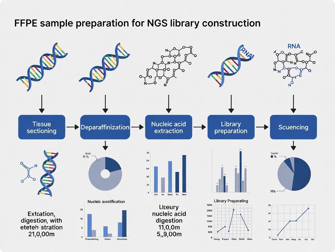
Mastering FFPE Sample Preparation for Robust NGS Library Construction: A Comprehensive Guide for Researchers
Next-generation sequencing (NGS) of Formalin-Fixed Paraffin-Embedded (FFPE) samples unlocks vast potential for cancer research and clinical diagnostics, yet the path to high-quality data is fraught with technical challenges.
Mastering FFPE Sample Preparation for Robust NGS Library Construction: A Comprehensive Guide for Researchers
Abstract
Next-generation sequencing (NGS) of Formalin-Fixed Paraffin-Embedded (FFPE) samples unlocks vast potential for cancer research and clinical diagnostics, yet the path to high-quality data is fraught with technical challenges. This article provides a comprehensive guide for researchers and drug development professionals, covering the foundational principles of FFPE-derived nucleic acid damage, modern methodological approaches for DNA and RNA library construction, advanced troubleshooting and optimization strategies, and a critical validation of different protocols and kits. By synthesizing the latest advancements and comparative data, this resource aims to empower scientists to reliably generate robust sequencing data from these precious but challenging archival samples, thereby accelerating discoveries in precision oncology.
Understanding FFPE Samples: Why They Are Challenging and Crucial for NGS
The FFPE Preservation Process and Its Impact on Nucleic Acids
Formalin-fixed paraffin-embedding (FFPE) is the cornerstone of tissue preservation in clinical and biomedical research, with an estimated 400 million to over 1 billion samples archived worldwide [1] [2]. While invaluable for pathological diagnosis, the chemical modifications inflicted upon nucleic acids present significant challenges for next-generation sequencing (NGS), potentially compromising variant detection accuracy and data reliability. Understanding these alterations and implementing robust mitigation strategies is therefore fundamental to unlocking the vast research potential of these archival resources. This application note details the impact of formalin fixation on DNA and RNA and provides optimized protocols to support successful NGS library construction from FFPE samples.
The FFPE Preservation Process and Induced Nucleic Acid Damage
The FFPE process involves tissue fixation in neutral buffered formalin, typically for 24 hours, followed by dehydration and embedding in paraffin wax for long-term storage at room temperature [3]. While ideal for morphological preservation, this process triggers multiple deleterious mechanisms that degrade nucleic acid quality.
Mechanisms of Formalin-Induced DNA Damage
Formalin fixation causes several types of chemical alterations to DNA, which can be classified into five key mechanisms [4]:
- Base Addition and Cross-linking: Formaldehyde reacts with nucleophilic amino groups on DNA bases, creating modified species with altered base-pairing abilities. These can further react to form methylene bridges, resulting in protein-DNA or DNA-DNA cross-links that block polymerase progression during amplification [4].
- Depurination and Strand Fragmentation: Formalin fixation accelerates the cleavage of glycosidic bonds, generating apurinic/apyrimidinic (AP) sites. These sites are highly susceptible to backbone cleavage under acidic or heated conditions, leading to pronounced DNA fragmentation [4].
- Base Deamination: Spontaneous deamination of cytosine to uracil (and 5-methylcytosine to thymine) is a frequently encountered artifact. This results in C>T/G>A base substitutions during sequencing, which can be misinterpreted as true variants, particularly in somatic cancer studies [4].
The following diagram illustrates the primary mechanisms of DNA damage caused by formalin fixation.
Comparative Quality of FFPE vs. Fresh Frozen Nucleic Acids
The cumulative effect of these damage mechanisms results in nucleic acids that are markedly inferior to those from fresh frozen (FF) tissue, the gold standard for NGS.
Table 1: Characteristic Differences Between FFPE and Fresh Frozen DNA
| Quality Metric | Fresh Frozen (FF) DNA | FFPE DNA | Experimental Consequence |
|---|---|---|---|
| A260/A280 Ratio | ~1.8 [3] | ~1.8 [3] | Purity is generally maintained in FFPE DNA. |
| A260/A230 Ratio | High (typically >2.0) | 0.9 ± 0.2 [3] | Indicates salt or solvent contamination, requiring rigorous purification. |
| DNA Integrity Number (DIN) | High (typically >7) | 5.5 ± 0.6 [3] | Direct measure of fragmentation; lower DIN correlates with reduced library complexity. |
| Average Fragment Size | >10,000 bp | ~7,573 bp [3] | Limits the size of amplifiable fragments and can bias sequencing coverage. |
| Primary Artifact Types | Low background | C>T/G>A substitutions, other single base changes [4] | Leads to false positive variant calls, requiring specialized bioinformatic filtering. |
Similar challenges affect FFPE-derived RNA, which is often highly fragmented. Metrics like the DV200 (percentage of RNA fragments >200 nucleotides) are used for quality assessment, with values >30-60% generally considered usable for sequencing, though with limitations [5] [6].
Pre-Analytical Quality Assessment and Mitigation Strategies
Rigorous quality control (QC) is the most critical step in ensuring successful NGS from FFPE samples.
DNA Quality Control Workflow
A comprehensive QC workflow assesses both physical degradation and chemical damage.
- Spectrophotometry (NanoDrop): Assesses DNA purity via A260/A280 and A260/A230 ratios. A low A260/A230 ratio in FFPE samples indicates the need for further clean-up [3].
- Fluorometric Quantification (Qubit): Provides accurate DNA concentration, superior to spectrophotometry for fragmented samples.
- Fragment Analysis (TapeStation, Bioanalyzer): Determines the DNA Integrity Number (DIN) or the distribution of fragment sizes, directly informing the expected insert size of NGS libraries [3].
- qPCR-based QC: A highly recommended method that quantifies the amount of amplifiable DNA, which is a better predictor of library prep success than fluorometry alone, as it reflects cryptic chemical damage [7].
DNA Repair Treatments
To mitigate damage, enzymatic repair mixes can be employed prior to library construction. These typically include:
- Uracil-DNA Glycosylase (UDG): Removes uracils resulting from cytosine deamination, significantly reducing C>T artifacts [4].
- Endonuclease IV / AP Lyase: Cleaves AP sites and repairs the resulting strand breaks.
- DNA Polymerase: Fills single-base gaps.
The effectiveness of a pre-library repair step is illustrated by its ability to generate data from even highly compromised samples, such as 13-year-old FFPE liver tissue with a DIN of 2.0 [4].
Optimized NGS Library Construction from FFPE DNA
Library preparation from FFPE DNA requires specific optimizations to handle low-input, fragmented, and damaged material.
Fragmentation Method Comparison
The choice of fragmentation method significantly impacts coverage uniformity and variant detection sensitivity.
Table 2: Performance Comparison of DNA Fragmentation Methods for FFPE WGS
| Fragmentation Method | Coverage Uniformity | Performance in GC-Rich Regions | SNP False-Negative Rate | Key Considerations |
|---|---|---|---|---|
| Mechanical Shearing (e.g., Sonication) | More uniform [8] | Superior performance [8] | Lower at reduced sequencing depth [8] | Lower sequence-specific bias; requires capital investment and causes sample loss [7]. |
| Enzymatic Fragmentation | Less uniform, prone to bias [8] | Reduced sensitivity [8] | Higher at reduced sequencing depth [8] | Scalable and automatable; modern kits are optimized to minimize artifacts for FFPE [7]. |
| Tagmentation (Tn5-based) | Varies by kit | Varies by kit | Varies by kit | Fast and efficient; sequence bias must be evaluated for FFPE applications [8] [2]. |
Detailed Protocol: Enzymatic Library Preparation for FFPE DNA
This protocol is adapted from the Watchmaker DNA Library Prep Kit with Fragmentation, which is optimized for challenging FFPE samples [7].
Objective: To construct high-complexity, sequencing-ready libraries from variable-quality FFPE DNA while minimizing the introduction of artifacts.
Materials and Reagents:
- DNA Library Prep Kit with Fragmentation (e.g., Watchmaker)
- FFPE DNA (1-200 ng)
- Size-selection Beads (e.g., SPRIselect)
- Adapter Oligos (with unique dual indices for multiplexing)
- Thermal Cycler
- qPCR Kit for Library Quantification
Procedure:
- Fragmentation and A-tailing:
- In a single tube, combine 5-200 ng of FFPE DNA with Fragmentation/A-tailing Master Mix.
- Incubate at 30°C for 3 minutes (mild conditions suitable for FFPE), then at 65°C for 30 minutes to inactivate the enzymes.
Adapter Ligation:
- Add Ligation Master Mix and unique dual index adapters directly to the fragmentation reaction.
- Incubate at 20°C for 15 minutes. This single-tube protocol minimizes sample loss.
Post-Ligation Cleanup:
- Purify the adapter-ligated library using size-selection beads.
- To tailor the mean insert size for sequencing economy, adjust the bead-to-sample ratio:
- 0.8X: Standard ratio, retains a broader size distribution.
- 0.65X - 0.5X: Retains longer fragments, increasing average insert size but reducing yield.
Library Amplification:
- Amplify the cleaned-up library with a high-fidelity PCR mix for 4-12 cycles, depending on input.
- Use P5/P7 primers compatible with your sequencing platform.
Final Purification and QC:
- Perform a final 1X bead cleanup.
- Quantify the final library yield by qPCR and assess the size distribution using a TapeStation or Bioanalyzer.
Critical Steps and Troubleshooting:
- Input DNA Mass: If library yield is low, increase input DNA mass to 100-200 ng to improve complexity [7].
- Post-Ligation Cleanup Ratio: If the library is too fragmented, use a lower bead ratio (e.g., 0.5X) to selectively retain longer fragments [7].
- Amplification Cycles: Use the minimum number of PCR cycles necessary to avoid skewing library complexity and amplifying duplicates.
The following flowchart summarizes this optimized library preparation workflow.
The Scientist's Toolkit: Essential Reagents and Solutions
Success in FFPE-NGS relies on a suite of specialized reagents and kits designed to overcome the inherent challenges of the sample type.
Table 3: Key Research Reagent Solutions for FFPE-NGS
| Item | Function | Example Application |
|---|---|---|
| Specialized FFPE DNA/RNA Kits | Maximize yield and quality during nucleic acid extraction from paraffin-embedded tissues. | Maxwell FFPE Plus DNA Kit, truXTRAC FFPE Total NA kits [3] [8]. |
| DNA Damage Repair Mix | Enzymatically reverses common FFPE artifacts (deamination, abasic sites) to reduce false positives. | PreCR Repair Mix, UDG treatment [4]. |
| FFPE-Optimized Library Prep Kits | Designed for fragmented, low-input DNA; often feature enhanced enzymatic fragmentation. | Watchmaker DNA Library Prep Kit with Fragmentation [7]. |
| FFPE-Tn5 Transposase | A modified transposase engineered to function efficiently on damaged, cross-linked FFPE DNA. | scFFPE-ATAC for single-cell chromatin accessibility [2]. |
| Targeted Sequencing Panels | Focus sequencing power on clinically relevant genes, ideal for low-quality/quantity FFPE inputs. | TruSight Oncology 500 (TSO500) [8] [9]. |
| Stranded RNA-Seq Kits | Enable transcriptome profiling from degraded FFPE RNA; some are optimized for very low input. | TaKaRa SMARTer Stranded Total RNA-Seq Kit, Illumina Stranded Total RNA Prep [5]. |
FFPE samples represent an unparalleled resource for biomedical research. A detailed understanding of the formalin-induced damage mechanisms—including fragmentation, cross-linking, and base deamination—enables researchers to implement effective countermeasures. Through rigorous pre-analytical quality control, judicious use of DNA repair enzymes, and the application of modern, optimized library preparation protocols, high-quality NGS data can be reliably generated from these precious archival samples. This empowers robust retrospective studies and maximizes the utility of the vast global repository of FFPE tissues.
Formalin-Fixed, Paraffin-Embedded (FFPE) samples represent an invaluable resource in biomedical research and clinical diagnostics, with vast archives of preserved tumor tissues and rare clinical cases offering a window into historical pathology and molecular signatures [10]. The FFPE process, developed in the late 19th century, was originally designed to conserve tissue cellular morphology and protein epitopes, enabling pathologists to stain histological sections for morphological and immunohistochemical analyses [4]. However, the very fixation and storage methods that make these specimens durable also introduce significant challenges for molecular analysis—DNA extracted from FFPE samples is often degraded, cross-linked, and heavily fragmented, making it difficult to generate high-quality libraries for next-generation sequencing (NGS) [10].
The chemical modifications inflicted upon DNA during formalin fixation and long-term storage pose substantial technical hurdles for accurate sequencing. These challenges include analytical sample preparation failure and FFPE-induced chemical modifications that can lead to incorrect base identification [4]. The consequences can be serious, particularly for detection of false positive variants which are especially problematic for variant-based signatures and for somatic mutations of lower variant allele frequency (VAF) in cancer specimens [4]. Understanding the specific nature of FFPE-induced damage is therefore crucial for developing effective countermeasures in NGS library construction.
Molecular Mechanisms of FFPE-Induced DNA Damage
Formalin fixation triggers a spectrum of chemical alterations to DNA through distinct mechanistic pathways. The process begins with local strand separation in AT-rich genomic regions, which then magnifies due to increased susceptibility to further modifications, creating a vicious cycle of damage accumulation [4].
Classification of Damage Mechanisms
FFPE-induced DNA damage can be classified into five primary mechanistic processes:
Chemical Addition Reactions: Formaldehyde reacts with nucleophilic groups such as amino groups of DNA bases, resulting in modified base species with altered base pairing abilities [4]. These modified bases can further react to form covalent cross-links with other nucleophilic groups via methylene bridges [4]. During sequencing library preparation, such modifications can locally alter base pairing characteristics, leading to the incorporation of non-complementary nucleotides in daughter strands or blockage of DNA polymerase during amplification [4].
Glycosidic Bond Cleavage: Formaldehyde fixation accelerates the cleavage of glycosidic bonds and the generation of apurinic/apyrimidinic (AP) sites within the double strand [4]. These AP sites are more susceptible to damage and fragmentation and can lead to incorporation of alternative nucleotides [4]. DNA polymerases generally have low bypass efficacies for such AP sites, meaning these molecules may not be amplified sufficiently for sequencing, resulting in reduced library complexity and information loss [4].
Polydeoxyribose Fragmentation: The cleavage of the DNA backbone into separate segments is widely observed in FFPE-DNA [4]. Samples fixed in unbuffered formalin are particularly sensitive to increased DNA degradation because under acidic conditions, AP-sites form more easily by hydrolysis of protonated purines [4].
Spontaneous Deamination: The most frequently encountered chemical alteration of FFPE-DNA is spontaneous deamination of cytosine [4]. In living cells, this damage is repaired by glycosylases, but these repair enzymes are inactivated by fixation, allowing deamination events to accumulate [4]. Deaminated cytosine results in uracil, which pairs with adenine instead of guanine; when cytosine is methylated (5-methylcytosine), deamination leads to thymine that also pairs with adenine. Both cases lead to the base pair alteration C>T/G>A [4].
Table 1: Primary Types of FFPE-Induced DNA Damage and Their Consequences
| Damage Type | Chemical Basis | Impact on Sequencing |
|---|---|---|
| Base modifications | Addition of formaldehyde to nucleophilic groups on DNA bases | Altered base pairing, incorporation of incorrect nucleotides during amplification |
| Cross-links | Covalent methylene bridges between bases or DNA-protein | Polymerase blockage, amplification failure, underrepresented regions |
| AP sites | Cleavage of glycosidic bonds leading to loss of bases | DNA fragmentation, difficulty in amplification, reduced library complexity |
| DNA fragmentation | Backbone cleavage through polydeoxyribose breakdown | Short fragment lengths, uneven coverage, challenges in library construction |
| Cytosine deamination | Hydrolytic deamination of cytosine to uracil | C>T/G>A false substitutions, erroneous variant calls |
Quantitative Analysis of Sequencing Artefacts
The consequences of formalin fixation manifest as distinctive artefact patterns in sequencing data. Analysis of a 13-year-old FFPE sample compared to case-matched fresh frozen (FF) tissue revealed a specific repertoire of potential artefacts [4]. The two most prevalent artefact types in FFPE-extracted DNA are C>T/G>A changes caused by cytosine deamination and C>A/G>T changes that mostly result from base oxidation [4]. Other single base substitution artefacts such as T>A/A>T and T>C/A>G changes also contribute significantly to the total artefact repertoire [4].
In comparative analyses, the highest increase observed was a 7-fold increase for C>T/G>A artefacts in FFPE-DNA compared to FF-DNA [4]. The distribution of artefact allele frequencies (AAF) shows some artefacts exceeding 10% in analysed samples, with particularly high AAFs located in regions of low sequencing coverage where many genomic fragments are severely damaged and not amplified [4]. Those genomic fragments that are less severely damaged may result in artefact-bearing sequences that become overrepresented, leading to high AAFs that may stem from various root causes including oxidation or sequencing errors [4].
Table 2: Frequency and Characteristics of FFPE Sequencing Artefacts
| Artefact Type | Relative Increase in FFPE vs. FF | Typical Allele Frequency Range | Primary Chemical Cause |
|---|---|---|---|
| C>T/G>A | 7-fold increase | Up to >10% AAF | Cytosine deamination to uracil |
| C>A/G>T | Significant increase | Up to >10% AAF | Base oxidation |
| T>A/A>T | Equally prevalent in old samples | Variable | Multiple mechanisms |
| T>C/A>G | Equally prevalent in old samples | Variable | Multiple mechanisms |
| Indel artefacts | Increased by order of magnitude | Varies by tumour type | PCR-related during library prep |
Large-scale analysis of whole genome sequencing data from the England's 100,000 Genomes Project, comparing 578 FFPE samples with 11,014 fresh frozen samples across multiple tumour types, has identified three distinct artefactual signatures: one known (SBS57) and two previously uncharacterised (SBS FFPE, ID FFPE) [11]. This analysis demonstrated that compared to FF-derived samples, FFPE-derived samples yielded data of poorer quality, with smaller insert sizes (391 base pairs vs. 477 base pairs; p < 0.0001) and a higher percentage of chimeric DNA fragments (0.51% vs. 0.26%; p < 0.0001), indicative of damaged DNA templates [11].
Mitigation Strategies and Experimental Protocols
Successful sequencing of FFPE-derived DNA requires integrated mitigation strategies addressing pre-analytical quality control, wet-lab processing, and bioinformatic correction. A comprehensive approach across these domains is essential for generating reliable data from compromised samples.
Pre-Analytical Quality Control
Quality assessment of input DNA is an invaluable tool in establishing and optimizing an FFPE library preparation workflow. While electrophoretic methods provide indication of DNA degradation, they offer limited insight into chemical damage such as crosslinking, deamination, or other base modifications that impede conversion of FFPE DNA into sequencing libraries [7]. Quantitative PCR (qPCR)-based methods are recommended to determine the amount of amplifiable DNA in a sample, with "quality scores" from such assays typically serving as good predictors of FFPE library prep outcomes [7].
The DNA integrity number (DIN) is a valuable metric for assessing FFPE sample quality. Studies have demonstrated that successful variant detection is possible even from samples with low DIN scores. For instance, research on ovarian cancer samples identified significant variants including a single base insertion in TP53 at 2.8% allele frequency and an 18 bp deletion in TP53 at 23% allele frequency in samples with DIN scores of 3.0 and 2.6 respectively [12]. This demonstrates that valuable data can be obtained from moderately to heavily degraded samples when appropriate protocols are followed.
DNA Repair Treatments
DNA repair prior to library preparation has become essential for overcoming FFPE-induced damage. Specialized repair reagents have been developed to address specific types of damage commonly found in FFPE samples [10]. These optimized enzyme mixtures are specifically formulated to repair common types of FFPE-induced DNA damage including cytosine deamination to uracil, nicks and gaps, oxidized bases, and 3′-end blockage [10]. It is important to note that most repair reagents cannot address fragmentation or DNA-protein crosslinking, which must be managed through other approaches [10].
In comparative experiments, FFPE DNA repair reagents have demonstrated significant improvements in library yield for low-quality FFPE samples, while showing minimal difference in high-quality samples, indicating that these reagents specifically benefit compromised DNA without affecting intact inputs [10]. The implementation of repair treatments enables reduced DNA input down to 50 ng while maintaining good depth of coverage, extending the utility of precious samples with limited material [12].
DNA Repair Workflow for FFPE Samples
Library Preparation Optimization
Library preparation from FFPE DNA requires specialized approaches to accommodate damaged templates. Enzymatic fragmentation solutions have been developed specifically for FFPE samples, offering consistent, tunable insert sizes independent of input amount or FFPE quality, while significantly mitigating molecular artifacts associated with the library construction process [7]. These systems utilize improved chemistry and flexible parameters to enable consistent fragmentation and control over FFPE library insert size, with single-tube protocols that limit sample loss, improve library complexity and sequencing metrics, and enable full automation [7].
Post-ligation cleanup ratios can be adjusted to optimize library characteristics for sequencing. Reducing the SPRI ratio from the standard 0.8X to 0.65X or as low as 0.5X favors retention of longer fragments, which can help compensate for the shorter mean insert sizes typically observed in FFPE libraries [7]. This approach can increase peak fragment size for libraries produced from 5 ng of low-quality FFPE DNA to levels comparable to those obtained from high-quality FFPE samples using standard ratios [7].
The choice between hybridization capture and amplicon-based enrichment significantly impacts data quality from FFPE samples. Hybridization-based capture approaches consistently outperform amplicon-based methods in uniformity of coverage, with most samples achieving >99% of bases covered at >20% of the mean, ensuring that all bases within a panel can be assessed confidently [12]. Additionally, hybridization-based capture allows removal of PCR duplicates which can obscure minor alleles present within a sample [12].
Library Preparation Method Comparison
Research Reagent Solutions for FFPE Analysis
The development of specialized reagents has dramatically improved the quality of data obtainable from FFPE samples. These solutions target specific aspects of FFPE-induced damage and enable researchers to extract reliable genomic information from even heavily compromised samples.
Table 3: Essential Research Reagents for FFPE DNA Analysis
| Reagent Type | Specific Function | Key Benefits | Application Notes |
|---|---|---|---|
| FFPE DNA Repair Mix | Repairs common FFPE-induced damage including cytosine deamination, nicks, oxidized bases, and 3′-end blockage [10] | Significantly improves library yield for low-quality samples; enables input down to 50 ng [10] [12] | Minimal impact on high-quality DNA; specifically benefits compromised samples |
| High-Efficiency Library Prep Kits | Enzymatic fragmentation with optimized ligation chemistry; some include integrated fragmentation/A-tailing [7] | Consistent, tunable insert sizes; reduced artifacts; single-tube protocol minimizes sample loss [7] | Enables automation; improves library complexity and sequencing metrics |
| Hybridization Capture Panels | Target enrichment via biotinylated probes and streptavidin pull-down [12] | Superior uniformity of coverage (>99% bases >20% mean coverage); enables PCR duplicate removal [12] | Outperforms amplicon-based methods for FFPE samples; essential for confident variant calling |
| Post-Ligation Cleanup Beads | Size selection through adjustable SPRI ratios [7] | Allows optimization of fragment size distribution; improves sequencing economy | Lower ratios (0.5X-0.65X) retain longer fragments from degraded samples |
| qPCR Quality Assessment Kits | Quantification of amplifiable DNA despite damage [7] | Predicts library prep success more accurately than electrophoretic methods | Provides "quality scores" correlating with sequencing outcomes |
Bioinformatic Correction of FFPE Artefacts
Bioinformatic approaches play a crucial role in distinguishing true biological variants from FFPE-induced artefacts. Large-scale analyses have enabled the development of specialized tools and metrics for quantifying and correcting FFPE-specific damage patterns.
The development of an "FFPEImpact" score that quantifies sample artefacts has provided researchers with a standardized metric for assessing data quality [11]. This approach characterizes rather than discards artefacts, identifying specific artefactual signatures including one known (SBS57) and two previously uncharacterised (SBS FFPE, ID FFPE) signatures [11]. Analytical advancements now enable the identification of clinically actionable variants, mutational signatures, and permit algorithmic stratification despite inferior raw sequencing quality from FFPE-derived data [11].
A critical consideration in bioinformatic processing of FFPE data is the approach to variant filtering. Previous attempts to filter variants with allelic fractions of 10% or less have been shown to exclude genuine mutations, including clinically actionable variants present at low variant allelic fractions (VAFs) [11]. In one study, 7.7% of PIK3CA and BRAF V600E mutations occurred at a VAF < 10% and would have been discarded using such filtering thresholds [11]. Instead, correlation of allelic frequency with relative cancer cell content provides a more reliable approach, as true mutations demonstrate this correlation while artefacts do not [11].
FFPE samples remain an invaluable resource for biomedical research, particularly in cancer genomics, biomarker discovery, and retrospective clinical studies [10]. The comprehensive characterization of FFPE-specific DNA damage—including fragmentation, cross-links, and base modifications—has enabled the development of sophisticated countermeasures across the entire NGS workflow. Through integrated approaches addressing pre-analytical quality control, wet-bench processing with specialized reagents, and bioinformatic correction, researchers can now reliably extract genomic information from samples that were once considered unsuitable for sequencing.
While fresh frozen-derived WGS data remains the gold standard, FFPE samples can be used for WGS when necessary using the analytical advancements developed in recent years [11]. This potentially democratizes whole cancer genomics to many healthcare settings worldwide that lack the infrastructure for frozen tissue preservation [11]. As technologies continue to advance, the gap between FFPE and fresh frozen sample quality will likely narrow further, unlocking the tremendous potential of archival tissue banks for discovery research and clinical applications.
Formalin-fixed paraffin-embedded (FFPE) samples are invaluable resources in biomedical research and clinical diagnostics, providing access to vast archives of tissue specimens with associated clinical data. However, the very fixation process that preserves tissue morphology introduces significant challenges for next-generation sequencing (NGS). The chemical modifications and degradation caused by formalin fixation and paraffin embedding result in a spectrum of sequencing artifacts, biases, and data quality issues that compromise genomic analyses. Understanding these artifacts is crucial for accurate interpretation of sequencing data from FFPE-derived nucleic acids.
The core of the problem lies in the fundamental chemistry of formalin fixation. Formaldehyde induces multiple types of DNA damage through distinct mechanistic processes: chemical addition reactions that create altered base species, covalent cross-links between nucleic acids and proteins, accelerated cleavage of glycosidic bonds generating apurinic/apyrimidinic (AP) sites, polydeoxyribose fragmentation, and spontaneous cytosine deamination [4]. These modifications collectively contribute to the artifactual observations in downstream sequencing applications, potentially leading to false biological conclusions and incorrect clinical interpretations.
Molecular Mechanisms and Their Sequencing Consequences
Types of FFPE-Induced DNA Damage and Resulting Artifacts
FFPE processing triggers multiple molecular pathways that damage DNA, each with distinct consequences for sequencing data quality and interpretation. The primary mechanisms include:
Cytosine Deamination: Spontaneous deamination of cytosine to uracil (or 5-methylcytosine to thymine) results in C>T/G>A base substitutions during sequencing [4]. This represents the most frequently encountered chemical alteration in FFPE-DNA, with studies demonstrating a 7-fold increase in C>T/G>A artifacts compared to fresh frozen samples [4]. Since cellular repair enzymes are inactivated during fixation, these artifacts accumulate and are particularly problematic for detecting true somatic mutations in cancer genomics.
DNA Fragmentation and Cross-linking: Formaldehyde fixation accelerates cleavage of glycosidic bonds, generating AP sites that lead to DNA backbone fragmentation [4]. Additionally, covalent cross-links form between DNA and proteins, as well as within DNA strands themselves [13]. This damage manifests as reduced library complexity in NGS, with non-uniform coverage and dropout of specific genomic regions, particularly in AT-rich areas [4]. The polydeoxyribose fragmentation results in shortened DNA fragments (typically 225-300 bp) that are suboptimal for standard WGS workflows designed for 360-480 bp fragments [14].
Oxidative Damage: Oxidation of guanine to 8-oxoguanine leads to G>T/C>A transversions during sequencing [13]. This represents the second most prevalent artifact type in FFPE-extracted DNA, though it occurs less frequently than deamination artifacts [4]. The combination of these different damage types creates a complex background of artifactual variants that complicates variant calling, particularly for low-frequency somatic mutations.
Table 1: Types of FFPE-Induced DNA Damage and Their Sequencing Consequences
| Damage Type | Chemical Mechanism | Primary Sequencing Artifacts | Impact on Data Quality |
|---|---|---|---|
| Cytosine deamination | Deamination of cytosine to uracil, 5-methylcytosine to thymine | C>T/G>A base substitutions | False positive SNVs, altered mutational signatures |
| DNA-protein cross-links | Covalent bonds between DNA bases and proteins | Region-specific sequencing dropouts | Reduced library complexity, coverage gaps |
| Oxidative damage | Oxidation of guanine to 8-oxoguanine | G>T/C>A transversions | False positive SNVs, especially in GC-rich regions |
| AP site formation | Cleavage of glycosidic bonds | Random base incorporation, sequencing blocks | Reduced amplification efficiency, coverage bias |
| Backbone fragmentation | Polydeoxyribose cleavage | Short DNA fragments | Limited library yield, alignment challenges |
Quantitative Impact on Variant Calling
The cumulative effect of FFPE-induced damage significantly impacts variant calling accuracy across different mutation classes. Analysis of matched FF-FFPE sample pairs demonstrates that FFPE processing results in a median 20-fold enrichment in artifactual calls across mutation classes [14]. The distribution of these artifacts varies substantially by variant type:
Single Nucleotide Variants (SNVs): FFPE-derived WGS data shows a median 2.0x increase in SNV calls compared to matched fresh frozen samples, with some samples exhibiting up to 152x more SNVs [14]. This dramatically lowers SNV calling precision to approximately 50% in FFPE samples. The elevated artifact burden particularly affects genome-wide tumor mutational burden (TMB) calculations, which show substantial inflation in FFPE samples (median: 10.28 mutations/Mb) compared to matched FF (median: 3.45 mutations/Mb) [14].
Insertions/Deletions (Indels): FFPE processing similarly increases artifactual indel calls, with a median 2.4x enrichment compared to fresh frozen samples and precision reduced to 62% [14]. The spectrum of indel artifacts shows particular enrichment in repeat-mediated deletions, complicating the detection of true frameshift mutations in microsatellite regions [14].
Structural Variants (SVs): While SV calling precision remains relatively high (median 80%) with consensus calling approaches, sensitivity is significantly compromised (57%) due to reduced coverage and mapping quality issues arising from shorter read fragments [14]. FFPE-specific limitations in SV detection include a 15x lower coverage at FF-specific SV loci and hyper-segmentation in copy number variant profiles [14].
The following diagram illustrates the relationship between FFPE damage types and their effects on sequencing data:
Impact on Biomarker Detection
Effects on Complex Genomic Biomarkers
The artifactual background generated by FFPE processing substantially impacts the detection and quantification of complex genomic biomarkers used in research and clinical decision-making:
Tumor Mutational Burden (TMB): While coding TMB remains relatively unaffected, genome-wide TMB shows significant inflation in FFPE samples (median: 10.28, range: 1.42–536.38) compared to matched fresh frozen samples (median: 3.45, range: 0.04–561.56) [14]. Without consensus calling approaches, coding TMB shows an average 7-fold elevation in FFPE samples, potentially leading to incorrect immunotherapy eligibility assessments [14].
Homologous Recombination Deficiency (HRD): The elevated artifact burden impairs accurate detection of HRD status. In validation studies, HRD scores in FFPE data fell below detection cutoffs for 7/7 cases by HRDetect and 4/7 cases by CHORD compared to matched fresh frozen samples, resulting in incorrect HRD classification [14]. This has significant implications for PARP inhibitor therapy selection.
Mutational Signatures: FFPE damage induces characteristic artifactual mutational signatures that can obscure true biological signatures. Specifically, 45/56 FFPE samples showed increased contribution of SBS37 (median proportion: 23.4%) compared to corresponding fresh frozen samples (12/56, median proportion: 3.6%) [14]. This signature enrichment can interfere with accurate signature extraction and assignment, particularly for signatures associated with DNA damage repair deficiencies.
Table 2: Impact of FFPE Artifacts on Key Cancer Biomarkers
| Biomarker | FFPE-Induced Artifacts | Clinical/Research Implications | Mitigation Strategies |
|---|---|---|---|
| Tumor Mutational Burden (TMB) | 2-7x inflation in mutation burden | False positive immunotherapy biomarkers | Consensus calling, coding region focus |
| Homologous Recombination Deficiency (HRD) | Reduced HRD scores below clinical thresholds | Incorrect PARP inhibitor eligibility | Machine learning correction (FFPErase) |
| Microsatellite Instability (MSI) | Enrichment in repeat-mediated indels | Altered MSI calling accuracy | Panel-based approaches, size threshold adjustment |
| Mutational Signatures | SBS37 signature enrichment | Obscured true biological signatures | Signature decomposition tools |
| Copy Number Alterations | Hyper-segmentation, increased noise | Impaired detection of focal amplifications/deletions | Smoothing algorithms, coverage normalization |
Spurious Mutation Signature Enrichment
Beyond SBS37 enrichment, FFPE damage alters the apparent contribution of multiple mutational signatures. The elevated C>T transitions characteristic of cytosine deamination can mimic aging-related signatures or obscure true signature activities. The combination of elevated genome-wide mutation burden and corresponding artifact signatures creates particular challenges for detecting composite mutation signatures like HRD that rely on specific patterns of small mutations and structural variants [14].
The consequences extend beyond single-base substitutions to indels and structural variants. FFPE-derived data exhibits a 2.8x increase in both insertions and repeat-mediated deletions [14], which can interfere with accurate microsatellite instability (MSI) detection. In contrast, SV profiles remain largely unaffected (median cosine similarity: 0.97 between FF and FFPE) [14], suggesting that SV-based biomarkers may be more robust to FFPE artifacts than SNV-based biomarkers.
Experimental Assessment Protocols
DNA Quality Control Framework
Implementing robust quality control measures is essential for assessing FFPE DNA suitability for sequencing applications. A comprehensive nanoscale quality control framework incorporating both gel electrophoresis and quantitative PCR provides critical assessment of DNA integrity:
Gel Electrophoresis Analysis: Standardized agarose gel electrophoresis (1% agarose gel, 100V for 60 minutes in TAE buffer) enables visual assessment of DNA fragmentation patterns [15]. High-quality FFPE DNA should show a smear concentrated in the 200-1000 bp range, while severely degraded samples display a concentration of fragments below 200 bp. Denaturing polyacrylamide gel electrophoresis (10% denaturing gel, 120V in TBE buffer) provides higher resolution assessment of fragment size distribution [15].
qPCR Amplification Efficiency: Single-plex qPCR amplification of targets of varying lengths provides a quantitative measure of DNA amplifiability [15]. The protocol utilizes a CFX96 Real-Time PCR Thermal System with reaction volumes of 10 μL comprising 5 μL of 2× SYBR Green master mix, 1 μL of 4 μM forward primer, 1 μL of 4 μM reverse primer, 2 μL of nuclease-free water, and 1 μL of extracted gDNA. Thermal cycling conditions include initial denaturation at 95°C for 2 minutes, followed by 40 cycles of denaturation at 95°C for 10 seconds and annealing/extension at 60°C for 30 seconds [15]. A quantifiable inverse correlation exists between the degree of DNA fragmentation and amplification efficiency in FFPE samples [15].
DV200 Assessment for RNA: For FFPE RNA applications, the DV200 value (percentage of RNA fragments >200 nucleotides) predicts sequencing success. Samples with DV200 values below 30% are generally too degraded for reliable RNA-seq, while values between 30-50% may require specialized library preparation methods, and values above 50% indicate good quality FFPE RNA [5].
The following workflow illustrates the recommended quality control process for FFPE samples:
Library Preparation Method Comparisons
Selection of appropriate library preparation methods significantly impacts data quality from FFPE samples. Recent comparative studies of FFPE-compatible stranded RNA-seq library preparation kits reveal important performance differences:
Input Requirements and Success Rates: The TaKaRa SMARTer Stranded Total RNA-Seq Kit v2 (Kit A) achieves comparable gene expression quantification to the Illumina Stranded Total RNA Prep Ligation with Ribo-Zero Plus (Kit B) while requiring 20-fold less RNA input [5]. This advantage is crucial for limited samples, though Kit A requires increased sequencing depth to compensate for higher rRNA content (17.45% vs. 0.1%) and duplication rates (28.48% vs. 10.73%) [5].
Gene Detection and Quantification: Despite methodological differences, both kits show high concordance in differential gene expression analysis, with 83.6-91.7% overlap in identified differentially expressed genes and nearly identical detection of genes covered by at least 3 or 30 reads [5]. Housekeeping gene expression levels show highly significant correlation between kits (R² = 0.9747, p-value < 0.001) [5].
Pathway Analysis Concordance: Enrichment analysis using KEGG database demonstrates that 16/20 up-regulated and 14/20 down-regulated pathways show consistent enrichment/depletion between the two kits, indicating that biological interpretation remains consistent despite technical differences [5].
For DNA sequencing, the NEBNext UltraShear FFPE DNA Library Prep Kit utilizes a specialized enzyme mix for DNA repair and fragmentation, demonstrating improved sequence complexity and coverage uniformity from FFPE-derived DNA [13]. The repair step specifically targets damaged bases while preserving true mutations, with the critical advantage that polymerase activity occurs after damaged base removal to prevent fixation of artifacts [13].
Mitigation Strategies and Solutions
Computational Artifact Correction
Advanced computational methods have been developed specifically to address FFPE-derived sequencing artifacts:
Consensus Calling Approaches: Implementing consensus variant calling using multiple variant callers significantly reduces artifactual calls, particularly for structural variants where FFPE-specific calls decrease by 98% (from 92% to 12%) [14]. However, this approach shows limited efficacy for SNVs and indels, where the median proportion of FFPE-specific mutations remains high (62% and 73% respectively) even after consensus calling [14].
Machine Learning Classification: The FFPErase framework employs a random forest classifier to filter SNV/indel artifacts and deliver clinical-grade variant reporting [14]. This approach demonstrates 99% sensitivity compared to FDA-approved panel tests while reporting 24% more clinically relevant findings, effectively bridging the quality gap between FFPE and fresh frozen WGS data [14].
Bioinformatic Filtering Strategies: Artifact allele frequency (AAF) thresholds can effectively filter many FFPE artifacts, particularly when set at 5% or higher [4]. However, high-AAF artifacts occurring in regions of low sequencing coverage remain challenging and require additional contextual filters [4].
Enzymatic Repair Methods
Enzymatic repair of FFPE DNA prior to library preparation significantly improves data quality:
Commercial Repair Kits: Specialized FFPE DNA repair reagents (e.g., Hieff NGS FFPE DNA Repair Reagent, PreCR repair mix) target specific damage types including cytosine deamination to uracil, nicks and gaps, oxidized bases, and 3′-end blockage [10] [15]. These enzyme mixtures demonstrate significant improvement in library yields for low-quality FFPE samples without affecting intact inputs [10].
Workflow Integration: Incorporating repair steps before fragmentation and amplification is critical for optimal artifact reduction [13]. The NEBNext FFPE DNA repair V2 mix selectively targets damaged DNA bases, excising damaged portions in single-stranded DNA and performing base excision repair on double-strand damage [13]. This approach prevents over-fragmentation, retains intact DNA, and preserves true mutations while removing artifactual bases.
Performance Validation: Comparative whole-exome sequencing analysis of endometrial carcinoma samples with different archival durations demonstrates that enzymatic repair strategies significantly reduce base substitution artifacts while improving amplification efficiency at previously underrepresented genomic sites [15].
The Scientist's Toolkit: Research Reagent Solutions
Table 3: Essential Reagents for FFPE Sequencing Studies
| Reagent/Kit | Primary Function | Key Applications | Performance Notes |
|---|---|---|---|
| Hieff NGS FFPE DNA Repair Reagent | Enzymatic repair of FFPE-induced damage | WGS, WES from FFPE DNA | Repairs deamination, nicks, oxidized bases; improves library yield [10] |
| NEBNext UltraShear FFPE DNA Library Prep Kit | Library preparation from FFPE DNA | WGS, target enrichment from challenging samples | Combines repair and fragmentation; automation-friendly [13] |
| PreCR Repair Mix | DNA damage repair | Restoration of amplifiable templates from degraded DNA | Addresses deaminated cytosines, oxidized guanine [15] |
| QIAamp DNA FFPE Tissue Kit | Nucleic acid extraction | DNA isolation from FFPE tissues | Standardized extraction for consistent yield [15] |
| TaKaRa SMARTer Stranded Total RNA-Seq Kit v2 | RNA library preparation | Transcriptomics from low-input FFPE RNA | Requires 20-fold less input than conventional methods [5] |
| Illumina Stranded Total RNA Prep Ligation with Ribo-Zero Plus | RNA library preparation | FFPE RNA-seq with ribosomal RNA depletion | Superior rRNA depletion (0.1% rRNA content) [5] |
FFPE specimens present significant challenges for next-generation sequencing due to the diverse artifacts and biases introduced during fixation and storage. The molecular consequences include elevated false positive variant calls, impaired detection of complex biomarkers, and substantial data quality issues that vary in severity across mutation classes. However, integrated experimental and computational approaches—including rigorous quality control, enzymatic repair methods, specialized library preparation protocols, and advanced bioinformatic correction—can effectively mitigate these artifacts. The continuing development of improved mitigation strategies promises to further enhance the utility of FFPE-derived sequencing data for both research and clinical applications, ensuring that these invaluable archival resources can continue to drive discoveries in cancer biology and precision medicine.
The Unmatched Value of FFPE Archives in Translational and Clinical Research
Formalin-Fixed Paraffin-Embedded (FFPE) tissue samples represent an invaluable resource in biomedical research, comprising over 90% of clinical pathology specimens archived worldwide [6]. These archives, containing vast collections of tissues with associated clinical and outcome data, provide an unparalleled foundation for translational research and the development of precision medicine strategies. The ability to leverage these samples for next-generation sequencing (NGS) has transformed our approach to understanding disease biology, particularly in oncology [16]. While FFPE samples present unique technical challenges due to nucleic acid fragmentation and cross-linking, recent advances in library preparation technologies and spatial transcriptomics have unlocked their potential for comprehensive genomic, transcriptomic, and epigenomic analyses [6] [5]. This application note details the methodologies and experimental protocols that enable researchers to extract maximum scientific value from these precious clinical resources, highlighting the critical role of FFPE archives in advancing clinical research and therapeutic development.
Applications and Performance Benchmarks
Comprehensive Genomic Profiling with Targeted NGS Panels
Targeted next-generation sequencing panels have emerged as powerful tools for comprehensive genomic profiling of FFPE-derived nucleic acids, enabling detection of critical biomarkers for therapy selection.
Table 1: Analytical Validation of a 1021-Gene NGS Panel for FFPE Tissues [17]
| Parameter | Performance Metric | Specifications |
|---|---|---|
| Variant Types | SNVs/Indels, CNVs, Fusions | All variant types detected |
| Sensitivity | 100% at 2% VAF, 84.62% at 0.6% VAF | >99% for SNVs/Indels |
| Specificity | 100% for all variant types | No false positives observed |
| Input Material | ≥50 ng DNA | FFPE tissue or liquid biopsy |
| Coverage | ≥500× for 2% VAF, ≥2000× for 0.5% VAF | 99% of targets covered at ≥50× |
| Quality Metrics | Fraction of base quality ≥Q30: 94.7% | High confidence base calling |
| TMB & MSI | Accurate detection | Immunotherapy biomarkers |
The clinical utility of this approach was demonstrated in a validation study of over 1300 solid tumor samples, which revealed actionable alterations in more than 50% of cases, with on-label treatment biomarkers identified in 12.57% of patients, increasing to 20.15% when immunotherapy markers were included [17].
Spatial Transcriptomics in FFPE Tissues
Imaging-based spatial transcriptomics (iST) platforms have overcome previous limitations to enable high-plex gene expression analysis directly in FFPE tissue sections while preserving spatial context.
Table 2: Benchmarking Performance of Commercial iST Platforms on FFPE Tissues [6]
| Platform | Chemistry Principle | Transcript Count | Cell Segmentation | Concordance with scRNA-seq |
|---|---|---|---|---|
| 10X Xenium | Padlock probes with rolling circle amplification | Consistently high | Improved with membrane staining | High concordance |
| Nanostring CosMx | Branch chain hybridization | Highest total recovery | Slightly more clusters than MERSCOPE | High concordance |
| Vizgen MERSCOPE | Direct hybridization with probe tiling | Lower than competitors | Fewer clusters than Xenium/CosMx | Varying degrees |
| Stereo-seq V2 | Random priming for total RNA capture | Enables immune repertoire | Single-cell resolution | Host-pathogen simultaneous profiling |
This benchmarking study, conducted on tissue microarrays containing 17 tumor and 16 normal tissue types, revealed that all three commercial platforms could perform spatially resolved cell typing with varying sub-clustering capabilities, with Xenium and CosMx finding slightly more clusters than MERSCOPE [6]. The random priming strategy employed by Stereo-seq V2 offers unbiased transcript capturing and uniform gene body coverage, increasing sensitivity to marker genes and efficiency of non-polyadenylated RNA profiling [18].
Whole Genome Sequencing from FFPE Material
Whole genome sequencing (WGS) from FFPE-derived DNA provides comprehensive genomic information beyond what is achievable with targeted panels, detecting complex biomarkers including mutational signatures and genome-wide copy number alterations.
Table 3: Performance of FFPE-Derived Whole Genome Sequencing in Metastatic Melanoma [16]
| Variant Type | Detection Rate vs. F1CDx | Clinical Utility |
|---|---|---|
| Somatic SNVs | 95% | Treatment guidance |
| Multinucleotide Variants | 98% | Clinical trial eligibility |
| Insertions/Deletions | 90% | Prognostic stratification |
| Amplifications | 76% | Therapeutic targeting |
| Homozygous Deletions | 96% | Resistance mechanism identification |
| Tumor Mutational Burden | R = 0.98 with F1CDx | Immunotherapy response prediction |
In a study of 78 metastatic melanoma samples, FFPE-derived WGS demonstrated robust analytical validity and suggested treatments or clinical trials for all cases, identifying additional markers in 38% and 71% of cases compared to FoundationOneCDx and a melanoma-specific panel, respectively [16].
Experimental Workflows and Methodologies
Nucleic Acid Extraction from FFPE Tissues
The initial and most critical step in FFPE sample processing is the extraction of high-quality nucleic acids, which requires optimized protocols to address fragmentation and cross-linking issues.
Diagram 1: FFPE Nucleic Acid Extraction Workflow
Protocol: Optimized Nucleic Acid Extraction from FFPE Tissues
- Sample Selection and Sectioning: Cut 5-10 μm thick sections from FFPE blocks using a microtome. For heterogeneous tissues, employ pathologist-guided macrodissection to enrich for regions of interest [5].
- Deparaffinization:
- Incubate sections with xylene (2 changes, 5 minutes each) to remove paraffin.
- Rehydrate through graded ethanol series (100%, 95%, 70% - 2 minutes each).
- Rinse with nuclease-free water.
- Proteinase K Digestion:
- Add proteinase K digestion buffer (1-2 mg/mL concentration).
- Incubate at 56°C for 3-16 hours (longer incubation may be needed for older samples) with gentle agitation [19].
- Nucleic Acid Extraction:
- For DNA: Use silica-based columns or magnetic beads optimized for FFPE samples.
- For RNA: Employ guanidinium thiocyanate-phenol-chloroform extraction or commercial FFPE RNA kits.
- Quality Control:
- Assess DNA/RNA concentration using fluorometric methods (Qubit).
- Evaluate fragmentation and quality using Bioanalyzer/TapeStation (DV200 > 30% for RNA) [5].
- Verify amplifiability through qPCR.
Library Preparation from FFPE-Derived Nucleic Acids
Library preparation from FFPE-derived material requires specialized approaches to address fragmentation, damage, and limited input material.
Protocol: DNA Library Preparation for FFPE Samples [20]
- DNA Repair Treatment:
- Treat 10-250 ng of FFPE DNA with repair enzymes to address formalin-induced damage.
- Incubate at appropriate temperature for 30 minutes.
- End Repair:
- Convert fragmented DNA into blunt-ended fragments using end repair enzyme mix.
- Incubate at room temperature for 15-30 minutes.
- Adapter Ligation:
- Employ specialized ligation chemistry (e.g., single-stranded ligation) to minimize chimera formation [20].
- Use unique molecular identifiers (UMIs) for error correction and accurate variant calling.
- Incubate with adapters for 15-30 minutes.
- Library Amplification:
- Perform limited-cycle PCR (6-10 cycles) with high-fidelity polymerase to minimize bias.
- Use unique dual indexes for sample multiplexing.
- Library Cleanup and QC:
- Purify using magnetic beads and quantify by qPCR.
- Assess size distribution using Bioanalyzer/TapeStation.
Protocol: RNA Library Preparation for FFPE Samples [5] [21]
- rRNA Depletion or Poly(A) Selection:
- For whole transcriptome analysis: Use ribosomal RNA depletion kits (Ribo-Zero Plus) to remove abundant rRNA.
- For mRNA sequencing: Employ oligo(dT) selection for polyadenylated RNA.
- cDNA Synthesis:
- Library Construction:
- Use strand-specific protocols to maintain strand orientation information.
- Incorporate UMIs for accurate quantification and duplicate removal.
- Amplification and QC:
- Perform limited-cycle amplification (10-15 cycles).
- Assess library quality and quantity before sequencing.
Method Selection for Transcriptomic Profiling
The choice between whole transcriptome and 3' mRNA sequencing approaches depends on research goals, sample quality, and project scope.
Diagram 2: RNA-Seq Method Selection Guide
Essential Research Reagents and Solutions
Table 4: Key Research Reagent Solutions for FFPE NGS Library Construction [20] [5] [22]
| Reagent Category | Specific Product Examples | Function and Application |
|---|---|---|
| Library Prep Kits | xGen cfDNA & FFPE DNA Library Prep Kit [20] | Specialized for fragmented DNA; enables low VAF detection |
| Library Prep Kits | Illumina DNA Prep [22] | Bead-linked transposome tagmentation for uniform coverage |
| RNA Library Kits | TaKaRa SMARTer Stranded Total RNA-Seq Kit v2 [5] | Low input requirement (20-fold less RNA); maintains library complexity |
| RNA Library Kits | Illumina Stranded Total RNA Prep with Ribo-Zero Plus [5] | Effective rRNA depletion; high alignment rates for FFPE RNA |
| Enzymes | xGen 2x HiFi PCR Mix [20] | Superior GC-bias performance; reduces PCR duplicates |
| Unique Molecular Identifiers | xGen UDI Adapters [20] | Error correction; accurate variant calling in low-VAF situations |
| Hybridization Capture | xGen Hybridization Capture Reagents [20] | Target enrichment for focused sequencing applications |
| RNA Preservation | RNase inhibitors and stabilization reagents | Maintain RNA integrity during extraction process |
FFPE tissue archives represent a cornerstone of modern translational research, providing an unparalleled resource for biomarker discovery, disease mechanism elucidation, and therapeutic development. The methodologies and protocols detailed in this application note demonstrate the robust capabilities of current NGS technologies to overcome historical challenges associated with FFPE-derived nucleic acids. As spatial transcriptomics, single-cell analyses, and multi-omics integration continue to evolve, the value of these extensive clinical archives will only increase, further bridging the gap between basic research and clinical application. The ongoing optimization of library preparation methods and analytical pipelines ensures that FFPE samples will remain indispensable in the era of precision medicine, enabling researchers to extract maximum insight from these precious biomedical resources.
Proven Protocols: Building High-Quality NGS Libraries from FFPE DNA and RNA
Within the context of a broader thesis on FFPE sample preparation for NGS library construction, the initial quality control (QC) of extracted nucleic acids represents the most critical determinant of downstream sequencing success. FFPE archives represent an invaluable resource for cancer research and drug development, but the formalin fixation process introduces cross-linking, fragmentation, and chemical modifications that degrade nucleic acid quality [23] [5]. Consequently, rigorous, standardized QC is not a mere formality but an essential gatekeeping step to conserve resources, ensure data reliability, and prevent the misinterpretation of biological signals. This application note details the essential QC metrics and methodologies for evaluating FFPE-derived DNA and RNA, providing researchers with a structured framework for sample assessment prior to NGS library construction.
Critical Quality Control Metrics for FFPE Nucleic Acids
The evaluation of FFPE-derived nucleic acids requires a multi-faceted approach, moving beyond simple concentration measurement to assess fragmentation, purity, and functional integrity. The metrics summarized in Table 1 provide a composite picture of sample quality and predict suitability for specific NGS applications.
Table 1: Essential Quality Control Metrics for FFPE DNA and RNA
| Metric | Description | Assessment Method | Interpretation for FFPE Samples |
|---|---|---|---|
| DV200 | The percentage of RNA fragments greater than 200 nucleotides [24]. | Automated Electrophoresis (e.g., Agilent Bioanalyzer/TapeStation) [24]. | ≥ 30%: Generally required for successful RNA-Seq [5]. Higher values indicate better preservation. |
| DNA/RNA Integrity Number (DIN/RIN) | Algorithmic assessment of nucleic acid integrity. | Automated Electrophoresis (e.g., Agilent Bioanalyzer). | Of limited utility for highly fragmented FFPE samples. DV200 is preferred for RNA. |
| Concentration | Quantitative measure of nucleic acid yield. | Fluorescent assays (e.g., Qubit). | Essential for input normalization. Does not reflect integrity. |
| Purity (A260/A280 & A260/A230) | Ratios indicating contamination from protein or solvents. | UV Spectrophotometry (e.g., NanoDrop). | Ideal A260/A280: ~1.8-2.0. Deviations suggest protein or chemical contamination. |
| Fragment Size Distribution | Visualization of the fragmentation profile. | Automated Electrophoresis or qPCR-based assays. | Confirms expected fragmentation. Critical for determining shearing requirements for DNA. |
| Library Preparation Success | Efficiency of converting nucleic acids to a sequencer-compatible library. | qPCR or capillary electrophoresis of the final library. | Measures the ultimate goal: a high-complexity, adapter-ligated library ready for sequencing [20]. |
For FFPE RNA, the DV200 metric is particularly crucial. It directly addresses the challenge of RNA fragmentation by quantifying the proportion of RNA molecules that are long enough to be informative in downstream sequencing applications [24]. Studies have shown that the RNA extraction methodology itself significantly impacts these QC metrics and subsequent sequencing results, including the fraction of uniquely mapped reads and the number of detectable genes [23]. Therefore, consistent application of the extraction and QC protocol is vital for comparative analyses.
Experimental Protocols for Quality Control Assessment
Protocol: Determining RNA DV200 using Agilent Automated Electrophoresis Systems
The following protocol is adapted from Agilent's technical overview for the 2100 Bioanalyzer system, a cornerstone technology for FFPE RNA QC [24].
I. Principle Automated electrophoresis systems separate RNA fragments by size, generating an electrophoretogram and a digital gel image. The accompanying software calculates the DV200 value by determining the percentage of the total RNA population that exists as fragments larger than 200 nucleotides.
II. Equipment & Reagents
- Agilent 2100 Bioanalyzer, 4200 TapeStation, or 5300 Fragment Analyzer system.
- Appropriate RNA assay kit (e.g., RNA Nano, RNA Pico).
- RNA Marker and Gel-Dye mix.
- Magnetic stirrer and IKA vortex mixer.
- Heating block or bath set to 70°C.
- DV200 assay configuration file (downloadable from Agilent for specific software revisions).
III. Step-by-Step Procedure
- Sample Preparation: Dilute RNA samples to a concentration within the linear range of the assay (e.g., 25-500 ng/µL for Nano assays).
- Gel-Priming: Load the gel-dye matrix into the appropriate well of the microchip. Ensure no air bubbles are introduced.
- Sample Loading: Pipette 5 µL of the RNA marker into the ladder well and 1 µL of each sample into the sample wells.
- Chip Run: Place the chip in the adapter and vortex for 1 minute at 2400 rpm. Immediately transfer to the Bioanalyzer and start the run.
- Data Analysis with DV200:
- For New Data: Ensure the DV200 assay file is imported into the software. The % of total value for the region defined from 200 nucleotides to the upper limit (e.g., 10,000 nt) is the DV200 value [24].
- For Existing Data: The DV200 calculation can be applied retroactively by opening the data file, navigating to the 'Assay Properties' tab, and importing the appropriate DV200 setpoints file (.xsy) [24].
IV. Data Interpretation A DV200 value of ≥ 30% is commonly used as a threshold for proceeding with standard RNA-seq library preparation protocols [5]. Samples with DV200 values below this threshold may require specialized, degradation-tolerant library prep kits or should be considered for exclusion.
Protocol: Comparative Analysis of Library Prep Kits for Low-Quality RNA
This protocol outlines the methodology for a kit comparison study, as described in Scientific Reports (2025), which is essential for validating workflows for challenging FFPE samples [5].
I. Principle To empirically determine the optimal RNA-seq library preparation kit for specific sample types (e.g., low-input, low-DV200 FFPE RNA) by comparing performance metrics such as gene detection, mapping rates, and technical noise between different commercial kits.
II. Equipment & Reagents
- Two or more FFPE-compatible stranded RNA-seq kits (e.g., TaKaRa SMARTer Stranded Total RNA-Seq Kit v2 "Kit A" and Illumina Stranded Total RNA Prep Ligation with Ribo-Zero Plus "Kit B") [5].
- High-quality nucleic acids from a defined FFPE source (e.g., tumor specimens, cell line references).
- Equipment for library QC (Qubit, Bioanalyzer/TapeStation).
- Illumina NovaSeq 6000 or equivalent NGS platform.
III. Step-by-Step Procedure
- Sample Selection & Pathologist-assisted Macrodissection: Select FFPE blocks with high tumor content. For transcriptomic studies, precisely dissect regions of interest to exclude non-relevant tissue [5].
- Nucleic Acid Extraction: Extract RNA using a silica-based or isotachophoresis-based procedure. Record yield, DV200, and purity (A260/A280) for all samples [23] [5].
- Library Preparation: Prepare libraries in parallel using the kits under comparison, strictly following manufacturers' protocols. For example, Kit A may require 5 ng total RNA, while Kit B may require 100 ng [5].
- Library QC & Sequencing: Quantify final libraries, check fragment size distribution, and pool at equimolar ratios. Sequence on an Illumina platform to a sufficient depth (e.g., >50M paired-end reads).
- Bioinformatic Analysis:
- Primary Metrics: Calculate the percentage of uniquely mapped reads, rRNA content, and duplication rate.
- Gene Expression Metrics: Determine the number of genes detected (e.g., covered by ≥3 reads) and the percentage of reads mapping to exonic, intronic, and intergenic regions.
- Concordance Analysis: Perform Principal Component Analysis (PCA), differential expression analysis, and pathway enrichment (e.g., KEGG) to assess technical reproducibility and biological concordance between kits [5].
IV. Data Interpretation The optimal kit is identified by a balanced trade-off between input requirements and data quality. For instance, one kit may excel with low inputs while another may offer superior rRNA depletion and lower duplication rates [5].
Workflow and Decision Pathway Visualization
The following diagram illustrates the logical pathway for the initial assessment and subsequent direction of FFPE samples based on QC results.
FFPE Sample QC and Decision Pathway
The Scientist's Toolkit: Essential Research Reagent Solutions
Selecting the appropriate reagents and kits is fundamental to navigating the challenges of FFPE-derived nucleic acids. The solutions listed in Table 2 are critical for ensuring successful NGS outcomes.
Table 2: Key Research Reagent Solutions for FFPE NGS Workflows
| Reagent / Kit | Function | Key Feature / Benefit |
|---|---|---|
| xGen cfDNA & FFPE DNA Library Prep Kit (IDT) [20] | Preparation of sequencing libraries from degraded DNA. | Novel ligase minimizes chimera formation; high conversion rates for low-input samples. |
| KAPA HiFi DNA Polymerase [25] | PCR amplification during library prep. | Minimizes GC-bias, providing uniform coverage across regions with varying GC content. |
| Illumina Stranded Total RNA Prep with Ribo-Zero Plus [5] | RNA-seq library prep from total RNA (including FFPE). | Effective ribosomal RNA (rRNA) depletion (e.g., ≤ 0.1% rRNA). |
| TaKaRa SMARTer Stranded Total RNA-Seq Kit v2 [5] | RNA-seq library prep from total RNA. | Ultra-low input requirement (e.g., 5 ng), crucial for limited samples. |
| miRNeasy FFPE Kit (Qiagen) [23] | Silica-based column extraction of total RNA from FFPE. | Commonly used method; performance compared in studies. |
| Ionic FFPE to Pure RNA Kit (Protocol B) [23] | Isotachophoresis-based extraction of RNA from FFPE. | Showed superior performance in sequencing metrics vs. some silica-based methods. |
The choice between these solutions depends on specific experimental needs. For DNA library prep, the xGen kit is engineered for high complexity from degraded inputs [20]. For RNA, the decision may hinge on the available input material, favoring the TaKaRa kit for very low yields, versus the Illumina kit for its exceptional rRNA depletion when sample is not limiting [5]. Furthermore, the RNA extraction method itself has been shown to significantly impact sequencing results, with method B (Ionic) and C (iCatcher) outperforming method A (miRNeasy) in one study, yielding more uniquely mapped reads and a greater number of detectable genes [23].
Within next-generation sequencing (NGS) workflows, the fragmentation of DNA is a critical first step that profoundly influences the quality and reliability of downstream data. This choice is particularly crucial when working with challenging sample types like Formalin-Fixed Paraffin-Embedded (FFPE) tissues, where DNA is often cross-linked and degraded [26]. The core decision for researchers lies in selecting between two principal fragmentation methodologies: enzymatic and mechanical shearing. This application note provides a detailed comparison of these techniques, grounded in recent experimental data, and offers structured protocols to guide optimization of NGS library construction from FFPE samples, a common requirement in clinical oncology and translational research.
Technical Comparison: Enzymatic vs. Mechanical Fragmentation
The choice between enzymatic and mechanical fragmentation involves balancing multiple factors, including workflow efficiency, data quality, and sample requirements. The table below summarizes the core characteristics of each method.
Table 1: Key Characteristics of Fragmentation Methods
| Feature | Mechanical Fragmentation | Enzymatic Fragmentation |
|---|---|---|
| Principle | Uses physical force (e.g., acoustic shearing) to break DNA [27]. | Uses enzymes (e.g., transposases, nucleases) to cleave DNA [27]. |
| Uniformity & Bias | Superior coverage uniformity; minimal GC-bias [26] [8]. | Pronounced coverage imbalances, particularly in high-GC regions [26] [8]. |
| Variant Detection | Lower SNP false-negative and false-positive rates, especially at lower sequencing depths [26]. | Potential for reduced sensitivity in high-GC regions, which are often clinically relevant [26]. |
| Workflow & Throughput | Can involve sample transfer, leading to potential loss; may be limited in parallel processing [27]. | Amenable to high-throughput and automated workflows; steps can be combined in a single tube [27] [28]. |
| Sample Input & Loss | Potential for material loss during transfers; not ideal for very low inputs [27]. | Minimal sample loss; recommended for limited or precious samples [27]. |
| Initial Investment | Requires capital expenditure for instrumentation (e.g., Covaris) [27]. | No special instruments required outside standard lab equipment [27]. |
Impact on Coverage Uniformity and GC Bias
Recent comparative studies highlight a significant performance difference between the two methods. An evaluation of four PCR-free whole genome sequencing (WGS) workflows—one mechanical and three enzymatic—demonstrated that mechanical shearing via Adaptive Focused Acoustics (AFA) yielded a more uniform coverage profile across different sample types (blood, saliva, FFPE) and across the GC spectrum [26] [8]. Conversely, enzymatic workflows exhibited more pronounced coverage imbalances, disproportionately affecting regions with high GC content [26] [8]. This bias is non-trivial, as many clinically relevant genes implicated in hereditary disease and oncology are located in high-GC regions. In an analysis of 504 genes from the TruSight Oncology 500 panel, uniform coverage provided by mechanical fragmentation was critical for accurate variant detection and minimizing false negatives [26].
Practical Workflow Considerations
For labs processing a large number of samples or those with limited starting material, enzymatic fragmentation presents distinct advantages. It is easily scalable and can be integrated into automated liquid handling systems, reducing hands-on time and improving reproducibility for high-throughput sequencing facilities [27] [28]. The ability to perform fragmentation, end-repair, and adapter ligation in a single tube reaction also minimizes sample loss, making it suitable for precious or low-input samples [27] [29]. In contrast, mechanical shearing requires dedicated instrumentation and can involve more sample handling, but provides consistent performance regardless of sample GC content [27].
Recommended Protocols for FFPE Samples
The following protocols are adapted from manufacturer guidelines and recent research for preparing NGS libraries from FFPE-derived DNA.
Protocol A: Mechanical Fragmentation with AFA
This protocol utilizes the Covaris truCOVER PCR-free Library Prep Kit and is designed to maximize coverage uniformity [26] [8].
- Step 1: DNA Extraction. Extract DNA from FFPE tissue sections using a dedicated kit, such as the truXTRAC FFPE Total NA Auto 96 Kit, to maximize yield and quality [8].
- Step 2: DNA Quantification and Quality Control. Accurately quantify the extracted DNA using a fluorescence-based method (e.g., Qubit). Assess the degree of fragmentation via agarose gel electrophoresis or bioanalyzer.
- Step 3: Mechanical Shearing. Dilute the DNA to the required volume in a Covaris microTUBE. Shear the DNA using a Covaris instrument with settings optimized for FFPE-DNA to achieve a target insert size of ~200-350 bp [8] [30].
- Step 4: Library Preparation. Transfer the sheared DNA to a fresh tube. Proceed with end-repair, dA-tailing, and adapter ligation using the truCOVER kit, following the manufacturer's instructions. This protocol is PCR-free [8].
- Step 5: Library QC. Purify the library and assess its quality and concentration using a bioanalyzer and qPCR.
Protocol B: Enzymatic Fragmentation Workflow
This protocol is based on the NEBNext Ultra II FS DNA Library Prep Kit, which integrates fragmentation and library preparation into a streamlined workflow [31] [29].
- Step 1: DNA Extraction. Extract DNA using a standard FFPE DNA extraction method.
- Step 2: DNA Quantification. Quantify DNA as in Protocol A.
- Step 3: Enzymatic Fragmentation and Library Prep. Set up a single-tube reaction containing the DNA sample, fragmentation enzyme mix, and end-repair/dA-tailing reagents. Incubate at the recommended temperature (e.g., 20-minute fragmentation at 25°C) [29].
- Step 4: Adapter Ligation and PCR. Without a cleanup step, directly add sequencing adapters and ligase to the same tube to minimize sample loss. For low-input FFPE samples, proceed with a limited-cycle PCR amplification (e.g., 10 cycles) to generate sufficient library mass for sequencing [31] [29].
- Step 5: Library QC. Purify the final library and quantify using bioanalyzer and qPCR.
Table 2: Performance Data from FFPE Library Preparations using Enzymatic Fragmentation (NEBNext Ultra II)
| FFPE Sample | DNA Input (ng) | Library Yields (ng) | % Mapped | % Mapped in Pairs | % Duplication | % Chimeras |
|---|---|---|---|---|---|---|
| Kidney Tumor | 17 | 132 | 91.5 | 96.1 | 0.48 | 3.0 |
| Lung Tumor | 20 | 232 | 90.1 | 94.9 | 0.42 | 4.1 |
| Liver Normal | 20 | 691 | 92.6 | 94.7 | 0.33 | 8.6 |
| Breast Tumor | 30 | 514 | 91.9 | 95.1 | 0.37 | 4.5 |
Data adapted from NEB documentation showing library performance metrics from various FFPE tissues [31].
The Scientist's Toolkit: Essential Reagents and Kits
Selecting the appropriate library preparation kit is foundational to success. The following table lists key commercial solutions and their properties.
Table 3: Key Research Reagent Solutions for DNA Library Preparation
| Product Name | Fragmentation Method | Key Features | Ideal for FFPE? |
|---|---|---|---|
| truCOVER PCR-free Library Prep Kit (Covaris) | Mechanical (AFA) | PCR-free; optimized for uniform coverage and minimal GC-bias [26] [8]. | Yes, with optimized extraction [8]. |
| NEBNext Ultra II FS DNA Library Prep Kit (NEB) | Enzymatic | Integrated fragmentation & end-repair; high yields from low inputs; suited for automation [31] [29]. | Yes, as demonstrated with tumor samples [31]. |
| Illumina DNA Prep | Enzymatic (Tagmentation) | Fast, 3-4 hour workflow; flexible input (1-500 ng) [32]. | Yes, for fragmented DNA. |
| xGen ssDNA & Low-Input DNA Library Prep Kit (IDT) | Enzymatic | Specialized for low-quality degraded DNA and single-stranded DNA; input as low as 10 pg [32]. | Yes, for highly degraded samples. |
Workflow Visualization
The following diagram illustrates the key decision points and steps in the two fragmentation workflows, highlighting their parallel paths and divergent characteristics.
The decision between enzymatic and mechanical fragmentation for FFPE NGS library prep is multifaceted. Mechanical shearing is the superior choice for applications where data fidelity and uniform coverage are paramount, such in clinical diagnostics and variant discovery in GC-rich regions. Enzymatic fragmentation offers compelling practical advantages for high-throughput environments, studies with limited sample input, or where budget constraints are a primary concern. The optimal path forward depends on a clear alignment of the method's strengths with the specific goals, sample constraints, and resources of the research project.
Advanced DNA Repair Steps to Mitigate Artifacts and Improve Yield
Formalin-fixed paraffin-embedded (FFPE) samples are invaluable resources for clinical and cancer research, yet they present significant challenges for next-generation sequencing (NGS) due to extensive DNA damage. The formalin fixation process introduces chemical modifications including DNA-protein crosslinks, base alterations, and DNA fragmentation, while subsequent paraffin embedding can cause further degradation through heat and dehydration [33]. These damages lead to two primary problems in sequencing: (1) significantly reduced library yields due to polymerase blockage at damaged sites, and (2) sequencing artifacts that manifest as false-positive variants in mutation analysis [4]. Without proper mitigation, these artifacts can severely compromise data integrity, particularly in cancer genomics where detecting low-frequency somatic variants is critical. This application note details advanced DNA repair strategies to overcome these challenges and enable reliable sequencing from even highly degraded FFPE samples.
Understanding FFPE-Induced DNA Damage
Types of DNA Damage in FFPE Samples
The chemical alterations in FFPE-DNA are complex and multifaceted, requiring specific repair approaches for successful sequencing library construction. The primary damage types include:
- Cytosine deamination: Conversion of cytosine to uracil, resulting in C>T/G>A transitions during sequencing—the most prevalent artifact in FFPE samples [4]
- Oxidative damage: Formation of 8-oxoguanine leading to G>T/C>A transversions [33]
- Abasic sites: Loss of nucleotide bases through glycosidic bond cleavage [4]
- DNA fragmentation: Backbone cleavage producing short, damaged DNA fragments [4]
- Methylene bridge crosslinks: Covalent bonds between DNA and proteins or between DNA strands [33] [34]
- Nicks and gaps: Single-stranded breaks with non-uniform ends [33]
Table 1: Major DNA Damage Types in FFPE Samples and Their Sequencing Consequences
| Damage Type | Chemical Basis | Primary Sequencing Artifact | Relative Frequency |
|---|---|---|---|
| Cytosine Deamination | C → U deamination | C>T / G>A transitions | High (7-fold increase vs. FF) [4] |
| Oxidative Damage | G → 8-oxoG formation | G>T / C>A transversions | Moderate [33] |
| Abasic Sites | Base loss | Polymerase blockage | High [4] |
| DNA Fragmentation | Backbone cleavage | Reduced library complexity | Universal [33] |
| Crosslinks | Methylene bridges | PCR amplification failure | Variable [34] |
Impact on Sequencing Data Quality
The cumulative effect of these damages profoundly impacts NGS data quality. Artifactual variant calls can reach allele frequencies exceeding 10% in regions of low coverage, making true somatic variant identification particularly challenging [4]. Additionally, library preparation from FFPE-DNA often results in elevated duplication rates, chimeric reads, and uneven coverage—all contributing to reduced library complexity and increased sequencing costs [33] [35]. Understanding these artifacts is essential for developing effective repair strategies.
Comprehensive DNA Repair Workflow
A systematic approach to FFPE-DNA repair addresses both the restoration of damaged bases and the structural integrity of DNA fragments. The optimal workflow incorporates sequential repair steps that mirror cellular DNA repair pathways.
Diagram 1: Comprehensive FFPE-DNA repair and sequencing workflow
Pre-Repair Quality Assessment
Before initiating repair, assess DNA quality using multiple metrics:
- Quantitative PCR (qPCR): Compare amplification of short (≤100 bp) versus long (≥300 bp) amplicons to determine degradation index [4]
- DNA Integrity Number (DIN): Calculate using Bioanalyzer or TapeStation; samples with DIN ≥3.2 may be suitable for repair, with optimized protocols achieving improvement from 3.2 to 7.2 [36]
- Fragment size distribution: Analyze using microfluidic electrophoresis; successful repair should preserve higher molecular weight fragments [36]
- UV spectrophotometry: Determine purity via A260/A280 and A260/A230 ratios [36]
Specialized DNA Repair Enzymatic Mix
Advanced repair formulations target specific damage types sequentially:
- End Repair System: T4 DNA polymerase and T4 polynucleotide kinase to restore 5' phosphates and create blunt ends from non-uniform fragments [33]
- Nick and Gap Repair: DNA ligase seals single-stranded breaks, while polymerases fill gaps using the complementary strand as template [33]
- Uracil Excision: Uracil-DNA glycosylase recognizes and removes uracils resulting from cytosine deamination, leaving abasic sites for subsequent processing [4]
- Oxidative Damage Repair: Formamidopyrimidine DNA glycosylase (Fpg) recognizes and excises 8-oxoguanine lesions [33]
- Abasic Site Cleavage: AP endonucleases incise the DNA backbone at abasic sites created by glycosylases [4]
Table 2: DNA Repair Enzymes and Their Functions in FFPE-DNA Restoration
| Enzyme Category | Specific Enzymes | Function in FFPE Repair | Key Considerations |
|---|---|---|---|
| Glycosylases | UDG, Fpg, hOGG1 | Recognizes and removes damaged bases | UDG treatment essential for reducing C>T artifacts [4] |
| Endonucleases | AP Endonuclease, Endonuclease IV | Cleaves backbone at abasic sites | Creates single-nucleotide gaps for polymerization [4] |
| Polymerases | T4 DNA Pol, Bst Polymerase | Fills gaps using undamaged strand | Must have DNA damage bypass activity [33] |
| Ligases | T4 DNA Ligase, Taq DNA Ligase | Seals nicks after repair | Requires ATP as cofactor [33] |
| Kinases | T4 PNK | Restores 5' phosphate groups | Essential for subsequent adapter ligation [33] |
Implementation Protocols
Optimized DNA Extraction and Repair Protocol
The following protocol, adapted from Singh et al. (2025) and NEB applications, maximizes DNA yield and integrity from limited FFPE tissue [36].
Materials:
- QIAamp DNA FFPE Tissue Kit or QIAamp DNA FFPE Advanced Kit (Qiagen)
- NEBNext UltraShear FFPE DNA Library Prep Kit (NEB #E6655) or equivalent
- Thermal cycler with heated lid
- Magnetic stand for bead cleanups
- Bioanalyzer/TapeStation for quality control
Procedure:
Sectioning and Deparaffinization
- Cut 2-3 sections of 10-20 μm thickness from FFPE block
- Add 1 mL xylene to 10 mg tissue, vortex, incubate 5 minutes at room temperature
- Centrifuge at full speed for 5 minutes, remove supernatant
- Wash with 1 mL 100% ethanol, centrifuge, air dry pellet
Proteinase K Digestion and DNA Extraction
- Resuspend tissue in 200 μL buffer ATL with 20 μL Proteinase K
- Incubate at 56°C overnight with agitation (16-24 hours)
- Incubate at 90°C for 1 hour to reverse formalin crosslinks
- Continue extraction per manufacturer's protocol
- Elute in 30-50 μL Buffer AE
DNA Damage Repair Reaction
- Assemble the following repair mixture:
- FFPE DNA (10-100 ng recommended)
- 5 μL Repair Buffer (10×)
- 2.5 μL NAD+ (100×)
- 2.5 μL ATP (100×)
- 1.5 μL UDG (10 U/μL)
- 1.5 μL Fpg (8 U/μL)
- 1.0 μL Endonuclease IV (50 U/μL)
- 1.0 μL DNA Ligase (30 U/μL)
- 1.0 μL DNA Polymerase (10 U/μL)
- Nuclease-free water to 50 μL
- Incubate at 37°C for 2 hours
- Purify using 1.8× volume magnetic beads, elute in 22 μL EB
- Assemble the following repair mixture:
Quality Control Assessment
- Quantify using Qubit dsDNA HS Assay
- Assess integrity via Bioanalyzer DNA Integrity Number (DIN)
- Verify fragment size distribution (optimal range: 200-500 bp)
This optimized protocol has demonstrated an 82% increase in DNA yield and improved DIN from 3.2 to 7.2 compared to standard extraction methods [36].
Integrated Repair and Library Preparation
For optimal results, consider integrated workflows that combine repair with library preparation:
Diagram 2: Integrated repair and library preparation workflow
The NEBNext UltraShear FFPE DNA Library Prep Kit exemplifies this approach, selectively targeting damaged bases while preserving true mutations through specialized enzyme mixes [33]. This integrated method demonstrates robust performance across input amounts from 1-200 ng, with library yields ranging from 132-691 ng from FFPE-DNA inputs of 17-30 ng [37].
Research Reagent Solutions
Table 3: Essential Reagents for Advanced FFPE-DNA Repair
| Product Name | Manufacturer | Primary Function | Key Features |
|---|---|---|---|
| NEBNext UltraShear FFPE DNA Library Prep Kit | New England Biolabs | Integrated repair & library prep | Specialized enzyme mix reduces artifacts; workflow for 1-200 ng input [33] |
| QIAamp DNA FFPE Advanced Kit | Qiagen | High-yield DNA extraction | Optimized for challenging samples; compatible with repair protocols [36] |
| Maxwell RSC Xcelerate DNA FFPE Kit | Promega | Automated extraction & repair | Instrument-based; consistent low degradation indices [34] |
| Infinium FFPE DNA Restoration Kit | Illumina | Array-compatible restoration | Repairs DNA for methylation & genotyping studies [38] |
| TruSight Oncology 500 | Illumina | Targeted pan-cancer assay | Works with low-quality FFPE; detects TMB & MSI [38] |
Quality Control and Validation
Performance Metrics for Repaired DNA
After repair, assess success using these quantitative metrics:
- Library Conversion Rate: Percentage of input DNA converted to sequenceable library; should exceed 60% for successful repair [33]
- Mapping Efficiency: Typically >90% for repaired FFPE-DNA versus <80% for untreated controls [37]
- Chimeric Read Rate: <5% indicates effective repair of single-stranded overhangs [33] [37]
- Duplicate Read Percentage: <10% suggests good library complexity [37]
- Coverage Uniformity: >80% of target regions covered at 0.2× mean coverage demonstrates even representation [33]
Bioinformatic Artifact Filtering
Despite optimal wet-lab repair, some artifacts may persist, requiring bioinformatic filtering:
- ArtifactsFinder: Custom algorithm that identifies and filters artifacts derived from inverted repeat sequences and palindromic sequences in enzymatically fragmented libraries [35]
- ERROR-FFPE-DNA Checklist: Standardized framework for reporting FFPE-specific parameters to ensure fitness-for-purpose in publications [4]
- UMI-based Error Correction: Incorporation of unique molecular identifiers to distinguish true variants from amplification artifacts [39]
Advanced DNA repair protocols transform challenging FFPE samples into viable genetic material for NGS applications. The sequential approach—addressing nicks and gaps, excising damaged bases, and synthesizing across lesion sites—significantly improves library yield while reducing sequencing artifacts. When combined with integrated library preparation methods and appropriate bioinformatic filtering, these techniques enable reliable mutation detection from even highly degraded FFPE material. As FFPE samples continue to be invaluable resources for retrospective cancer studies and biomarker discovery, implementing these robust repair strategies ensures maximal information recovery from these historically challenging specimens.
Formalin-fixed paraffin-embedded (FFPE) samples represent an invaluable resource for clinical and translational research, with an estimated 50 to 80 million samples stored globally that are suitable for next-generation sequencing (NGS) analysis [40]. These samples are accompanied by rich clinical data, including primary diagnosis, therapeutic regimen, and patient outcomes, making them particularly valuable for retrospective studies in the era of personalized medicine [41] [40]. However, the RNA extracted from FFPE tissues presents significant challenges for sequencing library construction due to fragmentation and chemical modifications introduced during the fixation process [41] [42].
The fixation process causes RNA fragmentation and the formation of methylene bridges that alter nucleic acid structure, while subsequent dehydration and storage lead to further degradation [42]. This degradation results in RNA that typically shows a median RNA Integrity Number (RIN) of approximately 2.5 and a DV200 (percentage of RNA fragments >200 nucleotides) of 48%, in stark contrast to fresh frozen tissue which typically has a RIN of 8.1 and DV200 of 97% [42]. These technical challenges necessitate specialized approaches for RNA library construction that can effectively handle degraded transcripts while efficiently depleting abundant ribosomal RNA (rRNA), which normally constitutes ≥90% of total RNA [43].
This application note examines current methodologies and provides detailed protocols for constructing high-quality RNA sequencing libraries from FFPE-derived RNA, with particular emphasis on handling degraded transcripts and optimizing rRNA depletion strategies.
Technical Challenges in FFPE RNA Sequencing
RNA extracted from FFPE samples exhibits several characteristics that complicate library construction and subsequent sequencing. The fragmentation pattern of FFPE RNA typically shows a broad peak at <200 bp, as visualized by electropherogram trace [43]. This fragmentation is compounded by chemical modifications that reduce the efficiency of molecular biology enzymes used in library preparation [41].
The standard poly(A) enrichment methods commonly used in RNA sequencing are particularly unsuitable for FFPE samples due to the loss of the 3' poly(A) tail through degradation [41]. Furthermore, certain functionally important mRNAs are naturally non-polyadenylated and would be missed entirely with poly(A) selection approaches [41]. These limitations have driven the development of rRNA depletion-based methods that preserve more information from the total RNA pool.
Data generated from FFPE RNA-seq (fRNA-seq) exhibits distinctive characteristics including high rates of transcript dropout (zero counts), high variance in transcript counts, and susceptibility to extreme values due to fragmentation artifacts [42]. These properties make downstream analytical challenges substantial and necessitate specialized statistical approaches for accurate interpretation.
Methodological Approaches for FFPE RNA Library Construction
rRNA Depletion Strategies
Several rRNA depletion methods are currently employed for FFPE RNA sequencing, each with distinct mechanisms and performance characteristics:
RNase H-mediated Depletion: This method hybridizes DNA probes to rRNA followed by RNase H digestion of the RNA-DNA hybrids. This approach has been validated for library construction from 25 ng to 1 μg of total RNA and demonstrates strong performance with low-quality RNA, particularly degraded FFPE RNA [41]. The KAPA, QIAGEN, and Vazyme kits evaluated in comparative studies utilize variations of this method [41].
Probe-based Magnetic Depletion: This technique captures rRNA using complementary DNAs coupled to paramagnetic beads, physically removing rRNA from the reaction mixture [41].
ZapR Enzyme Depletion: This approach first transcribes total RNA to cDNA, then uses ZapR enzyme to digest all rRNA:DNA hybrids. The TaKaRa kit employs this method and is specifically designed for low-input samples (5-50 ng total RNA) with chemical modifications [41].
The following diagram illustrates the decision pathway for selecting the appropriate library construction method based on sample characteristics and research objectives:
Library Construction Principles: 3' mRNA-Seq vs. Whole Transcriptome Sequencing
Two principal approaches dominate FFPE RNA library construction, each with distinct advantages and applications:
3' mRNA-Seq focuses sequencing reads on the 3' ends of polyadenylated transcripts using oligo(dT) primers to initiate reverse transcription. This approach does not require prior poly(A) enrichment or rRNA depletion, efficiently shortening workflow time and reducing costs [40]. Since sequencing reads are concentrated at the 3' end, this method reduces sequencing depth requirements and associated costs for data analysis and storage. However, it primarily captures polyadenylated transcripts and provides limited information about transcript isoforms or non-coding RNAs [40].
Whole Transcriptome Sequencing employs random primers to initiate cDNA synthesis, enabling coverage across the complete transcript body. This method requires prior rRNA depletion to prevent abundant ribosomal RNAs from dominating the sequencing library [40]. Whole transcriptome approaches provide uniform gene body coverage, enabling detection of alternative splicing, fusion genes, and non-coding RNAs, including long non-coding RNAs (lncRNAs) that may serve as important biomarkers in various pathological states [40].
Table 1: Comparison of 3' mRNA-Seq and Whole Transcriptome Sequencing Approaches for FFPE Samples
| Parameter | 3' mRNA-Seq | Whole Transcriptome Sequencing |
|---|---|---|
| Principle | Oligo(dT) priming at 3' end | Random priming across transcript |
| rRNA Depletion | Not required | Required |
| Input RNA | 10-100 ng | 10-1000 ng |
| Best Applications | Differential expression | Isoform detection, fusion genes, non-coding RNA |
| Transcript Coverage | 3' UTR focused | Uniform across transcript |
| Cost Factors | Lower sequencing depth | Higher sequencing depth |
| Poly(A) RNA Only | Yes | No |
| Detection of Non-coding RNA | Limited | Comprehensive |
Comparative Performance of Commercial Kits
Recent studies have directly compared the performance of various commercial kits for FFPE RNA library construction. A 2025 study compared the TaKaRa SMARTer Stranded Total RNA-Seq Kit v2 (Kit A) and Illumina Stranded Total RNA Prep Ligation with Ribo-Zero Plus (Kit B) [5]. Both kits generated high-quality data, with important distinctions: Kit A achieved comparable gene expression quantification to Kit B while requiring 20-fold less RNA input, a crucial advantage for limited samples, albeit with increased sequencing depth requirements [5].
An earlier comparative analysis of four FFPE RNA library preparation kits (KAPA, TaKaRa, QIAGEN, and Vazyme) revealed that the TaKaRa kit, which uses a different principle of rRNA depletion (ZapR enzyme digestion after cDNA synthesis), showed the highest library yields and exon percentage in unique mapping data for FFPE samples, despite having higher residual rRNA [41]. The gene expression profiles using the same kit showed high concordance between FF and FFPE samples (R = 0.96-0.98), demonstrating the reliability of within-kit comparisons [41].
Table 2: Performance Metrics of Commercial Kits for FFPE RNA Library Construction
| Kit | Input Range | rRNA Depletion Method | Key Advantages | Residual rRNA | Unique Mapping Rate |
|---|---|---|---|---|---|
| TaKaRa SMARTer | 5-50 ng | ZapR enzyme post-cDNA synthesis | Ultra-low input capability | Higher (17.45%) [5] | Lower [5] |
| Illumina Stranded Total RNA | 10-1000 ng | Enzymatic depletion (Ribo-Zero Plus) | Excellent rRNA depletion (0.1%) [5] | Lower (0.1%) [5] | Higher [5] |
| KAPA | 25-1000 ng | RNase H method | High consistency with FF samples (R=0.98) [41] | Moderate | Moderate |
| QIAGEN | 1-100 ng | Similar to RNase H method | High concordance with KAPA [41] | Moderate | Moderate |
Detailed Protocols for FFPE RNA Library Construction
rRNA Depletion and Random-Primed cDNA Synthesis (Low-Input Protocol)
This protocol utilizes the TaKaRa SMARTer approach with RiboGone rRNA depletion and is optimized for low-input, degraded RNA from FFPE samples [43]:
Materials:
- RiboGone - Mammalian Kit (Takara Bio, Cat. #634847)
- SMARTer Universal Low Input RNA Kit for Sequencing (Takara Bio, Cat. #634940)
- NucleoSpin Total RNA FFPE Kit (or equivalent FFPE RNA extraction system)
- Agilent 2100 Bioanalyzer with RNA 6000 Pico Chip
Procedure:
- RNA Extraction: Extract total RNA from curls of FFPE tissue using the NucleoSpin TotalRNA FFPE kit according to manufacturer's protocol, using lysis method B with a 75-minute incubation at 56°C and optional on-column DNase treatment.
- RNA Quality Assessment: Validate extracted RNA quality using an Agilent 2100 Bioanalyzer with RNA 6000 Pico Chip. Expect a broad peak at <200 bp, characteristic of degraded FFPE RNA.
- rRNA Depletion: Incubate 30 ng of extracted total RNA with RiboGone - Mammalian kit according to manufacturer's specifications. This step specifically removes 5S, 5.8S, 18S, and 28S nuclear rRNA sequences and 12S mitochondrial RNA sequences.
- cDNA Synthesis: Convert 8 µl of rRNA-depleted RNA to cDNA using the SMARTer Universal Low Input RNA Kit with 18 PCR cycles for double-stranded cDNA amplification (increased cycles compensate for the small amount and degraded nature of the RNA).
- Library Preparation: Add Illumina adapters and indices using a low-input library preparation kit (ThruPLEX DNA-Seq Kit recommended).
- Sequencing: Sequence on an Illumina platform with 1×50 bp or 2×75 bp reads, aiming for ~6 million reads per sample for initial quality assessment.
Expected Outcomes: This protocol typically reduces rRNA reads to 0.6% of total reads and identifies approximately 16,463 genes with RPKM ≥0.1 from breast carcinoma FFPE tissue [43].
Single-Cell RNA Sequencing from FFPE Heart Tissue
For single-cell applications from FFPE tissues, the following protocol adapted from Vanegas et al. (2025) provides a robust workflow [44]:
Materials:
- 10x Genomics Chromium Fixed RNA Profiling Kit
- Liberase TH
- gentleMACS Octo Dissociator with Heaters
- Pre-Separation Filters (30μm)
- Countess II FL Automated Cell Counter
Procedure:
- Sample Selection: Assess RNA quality of FFPE heart tissue samples and select based on DV200 value ≥30% and visual inspection of H&E-stained sections.
- Deparaffinization:
- Prepare two intact 50 μm sections from rehydrated tissue block and transfer to gentleMACS C Tube.
- Add 3 mL xylene and incubate at room temperature for 10 minutes.
- Carefully remove xylene without disrupting tissue scrolls.
- Repeat xylene wash twice.
- Perform ethanol series: 100% ethanol (3 mL, then 1 mL), 70% ethanol (1 mL), 50% ethanol (1 mL), incubating 30 seconds each.
- Add 1 mL nuclease-free water, incubate 30 seconds, then remove.
- Add 1 mL PBS and keep sample on ice.
- Tissue Dissociation:
- Remove PBS and add 2 mL enzyme mix (420 µL of 5 mg/mL Liberase TH + 1,680 µL RPMI medium).
- Place tube on gentleMACS Octo Dissociator with heater and run program 37CFFPE1 (~48 minutes).
- Centrifuge at 300 rcf for 1 minute and resuspend pellet in supernatant.
- Filter through 30 µm Pre-Separation Filter.
- Cell Processing:
- Centrifuge cell suspension at 850 rcf at 4°C for 5 minutes.
- Resuspend pellet in 0.5 mL chilled Quenching Buffer.
- Determine cell concentration using automated cell counter.
- Library Construction: Continue with Chromium Fixed RNA Profiling protocol per manufacturer's instructions (10x Genomics User Guide CG000527).
- Sequencing: Sequence on Illumina NovaSeq X Series with 25B Reagent Kit (300 Cycle), aiming for 15,000 read pairs per cell.
Research Reagent Solutions
Table 3: Essential Research Reagents for FFPE RNA Library Construction
| Reagent/Kits | Manufacturer | Primary Function | Sample Compatibility |
|---|---|---|---|
| SMARTer Universal Low Input RNA Kit | Takara Bio | cDNA synthesis from low-input, degraded RNA | 200 pg-10 ng RNA [43] |
| RiboGone - Mammalian | Takara Bio | Depletion of rRNA sequences (5S, 5.8S, 18S, 28S, 12S mtRNA) | Human, mouse, rat RNA (10-100 ng) [43] |
| Illumina Stranded Total RNA Prep | Illumina | Whole transcriptome library prep with enzymatic rRNA depletion | Human, mouse, rat, bacteria (10-1000 ng) [45] [46] |
| NucleoSpin Total RNA FFPE Kit | Macherey-Nagel | RNA extraction from FFPE tissues | FFPE tissue sections/curls [43] |
| Chromium Fixed RNA Profiling Kit | 10x Genomics | Single-cell RNA profiling from fixed cells | Fixed cells from FFPE tissue [44] |
| Liberase TH | Sigma-Aldrich | Tissue dissociation for cell isolation | Various FFPE tissues [44] |
Data Analysis Considerations
Analysis of fRNA-seq data requires specialized approaches due to its unique characteristics. The data typically follows a negative binomial distribution, similar to bulk and single-cell RNA-seq data, but with higher rates of transcript dropout and greater variance [42]. Tools specifically designed for fRNA-seq data, such as PREFFECT (PaRaffin Embedded Formalin-FixEd Cleaning Tool), employ probabilistic frameworks to adjust for technical and biological variables while imputing missing values [42].
For alignment, HISAT2 and STAR are commonly used tools, with HISAT demonstrating that unique mapping ratios, percentage of exons in unique mapping reads, and number of detected genes all decrease with decreasing quality of input RNA [41]. Unique molecular identifiers (UMIs) are particularly valuable for fRNA-seq as they enable error correction and improve quantification accuracy by reducing artifacts from PCR amplification and transcript fragmentation [40] [45].
The following diagram illustrates the complete workflow from sample preparation to data analysis:
Successful RNA library construction from FFPE samples requires careful consideration of extraction methods, rRNA depletion strategies, and library preparation approaches tailored to the specific characteristics of degraded RNA. rRNA depletion methods coupled with random-primed cDNA synthesis have emerged as the most robust approaches for comprehensive transcriptome coverage from FFPE materials. The selection between 3' mRNA-Seq and whole transcriptome sequencing should be guided by research objectives, with the former ideal for differential expression analysis and the latter necessary for isoform detection, fusion genes, and non-coding RNA discovery.
As technologies continue to evolve, newer methods including single-cell spatial transcriptomics on FFPE sections are further expanding the research potential of these valuable clinical archives [18]. By applying the optimized protocols and analytical frameworks described in this application note, researchers can reliably extract high-quality transcriptomic data from even challenging FFPE samples, enabling robust biomarker discovery and translational research.
Formalin-Fixed Paraffin-Embedded (FFPE) samples represent an invaluable resource for cancer genomics and retrospective clinical studies, with over one billion archival samples available worldwide [47]. However, the very process that preserves tissue architecture—formalin fixation—induces significant nucleic acid degradation, fragmentation, and chemical modifications that present substantial challenges for next-generation sequencing (NGS) library construction [14] [34]. Success in deriving meaningful genomic data from these specimens hinges on a carefully tailored approach that considers input mass, utilizes strategic automation, and selects appropriate sequencing platforms. This application note provides detailed protocols and data-driven guidance to optimize FFPE DNA library preparation for diverse research applications, enabling researchers to maximize the value of these precious clinical samples.
Quantitative Foundations: Input Mass and Performance Metrics
DNA Input Mass Guidelines for FFPE Library Preparation
Table 1: Recommended DNA Input Mass Based on Sample Quality and Application
| Sample Quality | DNA Input Range | Recommended Applications | Key Considerations |
|---|---|---|---|
| High Quality (DIN >7) | 100-250 ng [20] | Whole Genome Sequencing, Whole Exome Sequencing | Maximizes library complexity and coverage uniformity |
| Moderately Degraded (DIN 4-7) | 50-100 ng [20] | Targeted Sequencing, Hybrid Capture | Balance between yield and data quality; may require additional PCR cycles |
| Severely Degraded (DIN <4) | 1-50 ng [20] | Low-pass WGS, Small Amplicon Panels | Ultra-low input protocols essential; higher PCR cycles needed; UMI incorporation critical |
Performance Metrics Across Input Mass and Quality
Table 2: Expected Performance Metrics from Optimized FFPE Protocols
| Parameter | Standard Protocol | Optimized Protocol | Improvement | Assessment Method |
|---|---|---|---|---|
| DNA Yield | Baseline | 82% increase [36] | Significant | NanoDrop 2000, Qubit dsDNA Assay [36] |
| DNA Integrity | DIN 3.2 [36] | DIN 7.2 [36] | 125% improvement | Bioanalyzer, TapeStation [36] |
| VAF Accuracy | ≤1% [20] | ≤1% [20] | High sensitivity maintained | Variant Allele Frequency measurement [20] |
| Artifact Reduction | 20-fold enrichment vs FF [14] | 98% reduction (SVs) [14] | Dramatic improvement | Consensus calling, FFPErase filtering [14] |
Experimental Protocols
Optimized DNA Extraction from FFPE Tissues
Principle: Maximize DNA yield and integrity while reversing formalin-induced cross-links and minimizing artifacts.
Reagents and Equipment:
- QIAamp DNA FFPE Tissue Kit or QIAamp DNA FFPE Advanced Kit (Qiagen) [36]
- Maxwell RSC Xcelerate DNA FFPE Kit (Promega) [34]
- Proteinase K
- Xylenes or other deparaffinization reagents
- Ethanol (96-100%)
- Nanodrop 2000 Spectrophotometer or equivalent
- Qubit Fluorometer with dsDNA HS Assay Kit
- Bioanalyzer or TapeStation system
Procedure:
- Sectioning: Cut 4-10 μm sections from FFPE block using a microtome. For limited tissue, use full scrolls. [36]
- Deparaffinization:
- Add 1 mL xylenes to samples, vortex, incubate 10 minutes at room temperature.
- Centrifuge at full speed for 5 minutes, carefully remove supernatant.
- Repeat once with xylenes, then twice with 96-100% ethanol. [34]
- Proteinase K Digestion:
- Resuspend pellet in 200 μL buffer containing 20 μL Proteinase K.
- Incubate at 56°C overnight (12-16 hours) with agitation. [34]
- Cross-link Reversal: Incubate at 90°C for 30-60 minutes. [36]
- DNA Purification: Continue with kit-specific protocol (Qiagen or Promega). [36] [34]
- Elution: Elute in 30-50 μL nuclease-free water or TE buffer.
- Quality Assessment:
Automated Library Preparation for FFPE DNA
Principle: Generate sequencing-ready libraries from FFPE DNA with minimal hands-on time while maintaining complexity.
Reagents and Equipment:
- xGen cfDNA & FFPE DNA Library Prep Kit (IDT) [20]
- xGen UDI Primers (IDT) [20]
- IntegenX Apollo 324 robot or Caliper Sciclone G3 [48]
- Thermal cycler
- Magnetic stand
Procedure:
- End Repair:
- Prepare reaction mix: 1-250 ng FFPE DNA, End Repair Enzyme Mix, reaction buffer.
- Incubate at 20°C for 30 minutes. [20]
- Ligation 1:
- Add Ligation 1 Adapter and Ligation 1 Enzyme directly to end repair reaction.
- Incubate at 25°C for 30 minutes.
- Note: This novel enzyme prevents adapter-dimer formation and chimera formation. [20]
- Ligation 2:
- Add Ligation 2 Adapter and appropriate enzymes.
- Incubate at 25°C for 30 minutes.
- The adapter gap-fills complementary bases to create double-stranded product. [20]
- Purification: Clean up ligation reaction using magnetic beads according to manufacturer's protocol.
- PCR Amplification:
- Set up PCR reaction: purified ligation product, xGen 2x HiFi PCR Mix, xGen UDI Primers.
- Cycle conditions: 98°C for 2 min; 7-14 cycles of 98°C for 15 sec, 60°C for 30 sec; 72°C for 1 min.
- Critical: Use minimum PCR cycles needed to maintain complexity (typically 7-10 for 50 ng input). [20]
- Final Purification: Clean up PCR product with magnetic beads, elute in 20-30 μL TE buffer.
- Quality Control:
- Quantify using Qubit dsDNA HS Assay.
- Assess size distribution using Bioanalyzer or TapeStation (expected peak: 200-500 bp).
- Automation Option: For high-throughput applications, implement on IntegenX Apollo 324 or Caliper Sciclone G3. [48]
Workflow Visualization
The Scientist's Toolkit: Essential Research Reagents
Table 3: Key Reagents for FFPE NGS Library Construction
| Reagent/Kit | Manufacturer | Function | Key Features |
|---|---|---|---|
| QIAamp DNA FFPE Kit | Qiagen [36] | DNA purification from FFPE tissues | Optimized for cross-link reversal; improved yield and integrity |
| xGen cfDNA & FFPE Library Prep Kit | IDT [20] | Library preparation from degraded DNA | Single-stranded ligation; minimal chimera formation; UMI compatibility |
| Maxwell RSC Xcelerate DNA FFPE Kit | Promega [34] | Automated DNA extraction | Rapid protocol; consistent yields; suitable for low-input samples |
| xGen 2x HiFi PCR Mix | IDT [20] | Library amplification | Superior GC-bias; high fidelity; reduced duplicates |
| xGen UDI Primers | IDT [20] | Library indexing | Unique dual indexes; reduce index hopping in multiplexed sequencing |
| Proteinase K | Various [34] | Protein digestion | Degrades cross-linked proteins; releases nucleic acids |
| xGen Hybridization Capture Reagents | IDT [20] | Target enrichment | Compatible with FFPE libraries; high on-target rates |
Analytical Framework and Quality Control
Quality Control Checkpoints
Post-Extraction QC:
- Quantity: Use fluorometric methods (Qubit) for accurate DNA quantification. [48]
- Purity: Assess A260/A280 ratios (target: 1.8-2.0) and A260/230 ratios (target: >2.0) via spectrophotometry. [48] [49]
- Integrity: Determine DNA Integrity Number (DIN) via Bioanalyzer/TapeStation. DIN >5 preferred for WGS, DIN >3.5 acceptable for targeted sequencing. [36]
Post-Library QC:
- Size Distribution: Verify library fragment size (typically 200-500 bp) via Bioanalyzer. [48]
- Concentration: Quantify using qPCR-based methods for accurate molarity determination. [48]
- Adapter Dimer Contamination: Check for primer-dimer peaks at ~120-150 bp. [49]
Computational Quality Assessment
FASTQ Quality Metrics:
- Utilize FastQC for per-base sequence quality assessment. [49]
- Q-score >30 indicates high-quality data for most applications. [49]
- Perform adapter trimming with CutAdapt or Trimmomatic before alignment. [49]
Variant Validation:
- For WGS applications, implement consensus calling to reduce FFPE-specific artifacts (98% reduction in artifactual SVs). [14]
- Apply specialized tools (FFPErase) for SNV/indel artifact filtration. [14]
- Monitor mutation signatures; SBS37 enrichment may indicate FFPE artifacts. [14]
Successful NGS library construction from FFPE specimens requires meticulous attention to input mass, strategic implementation of automation, and careful platform selection based on DNA quality and research objectives. The optimized protocols presented here demonstrate that with appropriate methodologies, DNA yield can be increased by 82% and integrity significantly improved, making even severely degraded specimens viable for genomic analysis. As FFPE samples continue to be invaluable for cancer research and biomarker discovery, these tailored approaches ensure maximum information recovery from these challenging yet precious clinical resources.
Solving Common FFPE NGS Pitfalls: From Low Yield to Sequence Artifacts
Diagnosing and Overcoming Low Library Yield and Poor Complexity
Within the context of FFPE sample preparation for Next-Generation Sequencing (NGS), achieving high library yield and complexity is a fundamental prerequisite for successful downstream genomic analyses. However, the very nature of formalin-fixed paraffin-embedded (FFPE) tissues often leads to suboptimal results, characterized by low library yield and poor complexity. These issues can severely compromise data quality, resulting in insufficient sequencing coverage, biased representation of genomic regions, and reduced variant-calling accuracy [50] [51]. This application note provides a detailed diagnostic framework and robust experimental protocols to overcome these challenges, ensuring the generation of high-quality sequencing libraries from even the most compromised FFPE samples.
The core challenges stem from the FFPE process itself. Formalin fixation induces DNA fragmentation, cross-links between nucleic acids and proteins, and various forms of base damage, such as cytosine deamination and oxidative damage [50] [51]. Consequently, DNA extracted from FFPE samples is often highly degraded, yielding limited amounts of fragmented nucleic acids with non-uniform ends. During library preparation, these damaged DNA molecules can lead to polymerase blockage, inefficient adapter ligation, and the formation of chimeric reads, ultimately manifesting as low library yield and poor complexity in sequencing data [50].
Diagnosis: Assessing Sample Quality and Library Metrics
Accurate diagnosis of DNA quality and the root causes of library failure is the critical first step. The following methods and metrics form the cornerstone of a reliable quality control (QC) pipeline.
Pre-Library Preparation Quality Control
Before committing valuable samples to library prep, perform these essential QC checks on the extracted FFPE DNA:
- Quantification using Fluorometric Methods: Use Qubit fluorometry for accurate DNA quantification. Avoid UV-spectrophotometry (e.g., Nanodrop), as it is prone to overestimation due to residual RNA, single-stranded DNA, and other contaminants [51].
- Assessment of DNA Amplifiability with qPCR: The Infinium FFPE QC Kit (or equivalent qPCR assay) is recommended to determine the fraction of amplifiable DNA. This assay calculates a ΔCq value, which is a strong predictor of library preparation success. A ΔCq value of ≤ 5 indicates good quality DNA, while a ΔCq > 5 suggests significant damage and predicts potential library failure or poor performance [52].
- Fragment Size Analysis: Utilize the Agilent Bioanalyzer or TapeStation to determine the DV200 value (the percentage of RNA fragments > 200 nucleotides) for RNA, or to assess the DNA fragment size distribution. Highly degraded DNA will show a pronounced smear at lower molecular weights.
Table 1: Quality Control Methods for FFPE-Derived Nucleic Acids
| Method | Metric | Interpretation of Results | Recommendation for Library Prep |
|---|---|---|---|
| Qubit Fluorometry | DNA/RNA Concentration (ng/µL) | Accurate quantification of double-stranded nucleic acids. | Use for input normalization. |
| qPCR (e.g., Infinium FFPE QC Kit) | ΔCq value | ΔCq ≤ 5: Good quality. ΔCq > 5: Highly degraded. | For DNA: If ΔCq > 5, use specialized repair protocols. [52] |
| Bioanalyzer/TapeStation | DV200 (for RNA); DNA Integrity Number (DIN) or fragment profile | DV200 > 55% for RNA: Good. Lower values indicate degradation. | For RNA: Adjust input amount based on DV200. [52] |
Post-Library Preparation Quality Control
After library construction, evaluate the following sequencing metrics to diagnose low yield and complexity:
- Library Yield: Quantified in nanograms of final library, a low yield often indicates inefficient adapter ligation or amplification, frequently caused by damaged DNA ends or insufficient input [53].
- Percentage of Duplicate Reads: A high duplication rate is a direct indicator of poor library complexity, meaning the sequencing library contains an insufficient number of unique DNA molecules. This is a common consequence of low input or extensive PCR amplification of a limited starting material [5].
- Mapping Rates and Chimeric Reads: A low percentage of reads mapped to the reference genome, or a high percentage of chimeric reads, can signal the presence of cross-linked or damaged DNA fragments that anneal inappropriately during library prep [53] [50].
Table 2: Key NGS Metrics for Diagnosing Library Issues
| Metric | Definition | Indicator of Problem |
|---|---|---|
| Library Yield | Mass of final library (ng) | Low yield indicates inefficiencies in ligation/amplification. |
| % Duplication | Percentage of mapped sequence that is marked as duplicate. | High percentage indicates poor library complexity. [53] [5] |
| % Mapped in Pairs | Percentage of reads whose mate pair was also aligned. | Low percentage suggests high fragmentation or damage. [53] |
| % Chimeras | Percentage of reads mapping to different chromosomes or outside max insert size. | High percentage suggests DNA crosslinking or annealing of single-stranded overhangs. [53] [50] |
The following diagram illustrates the logical workflow for diagnosing and troubleshooting low yield and poor complexity.
Overcoming the Challenges: Protocols and Reagents
A Robust DNA Repair and Fragmentation Workflow
Specialized library prep kits that integrate DNA repair mechanisms are highly effective for FFPE samples. The following protocol, based on the NEBNext UltraShear FFPE DNA Library Prep Kit, is designed to mitigate damage and improve outcomes [50].
Protocol: DNA Repair and Fragmentation for FFPE Samples
Principle: This workflow prioritizes the repair of DNA damage before fragmentation and library construction. This step excises damaged bases, fills in nicks and gaps, and removes single-stranded overhangs, which prevents the introduction of sequencing artifacts and boosts library conversion rates [50].
Research Reagent Solutions:
- NEBNext UltraShear FFPE DNA Library Prep Kit (NEB #E6655): A specialized kit containing all necessary enzymes and buffers for repair, fragmentation, and subsequent library construction.
- NEBNext FFPE DNA Repair V2 Mix: A proprietary enzyme mix that selectively targets and excises common FFPE-induced damage like cytosine deamination and oxidative lesions.
- Magnetic Bead-Based Cleanup Beads (e.g., SPRI beads): For post-reaction cleanups and size selection.
- Qubit dsDNA HS Assay Kit: For accurate quantification of input DNA and final library.
- Agilent Bioanalyzer High Sensitivity DNA Kit: For quality assessment of the final library.
Methodology:
DNA Repair Reaction
- Input DNA: 1-100 ng of FFPE-derived DNA. The protocol is designed to be largely sample quality-agnostic.
- Reaction Setup: Combine DNA with NEBNext FFPE DNA Repair V2 Mix and the supplied reaction buffer.
- Incubation: Incubate at 37°C for 30 minutes. This step removes damaged bases and repairs nicks and gaps, preventing over-fragmentation in the next step and preserving intact DNA molecules [50].
- Enzyme Inactivation: Heat-inactivate the repair enzymes at 70°C for 10 minutes.
Controlled Enzymatic Fragmentation
- Reaction Setup: Add the NEBNext UltraShear Fragmentase to the repaired DNA.
- Incubation: Incubate at 37°C for a defined period (e.g., 10-15 minutes). The time-dependent nature of this enzymatic fragmentation allows for consistent sizing without the risk of over-fragmentation, a key concern with pre-degraded FFPE DNA [50].
- Cleanup: Purify the fragmented DNA using magnetic beads.
Library Construction
- Proceed with standard library construction steps, including end-repair, dA-tailing, and adapter ligation, as per the kit's instructions. The prior repair and fragmentation steps ensure these downstream enzymatic reactions proceed with high efficiency.
Library Amplification & QC
- PCR Amplification: Amplify the library using a high-fidelity PCR master mix. The number of cycles should be optimized based on input.
- Example: For low input (17-50 ng), 10-12 cycles may be sufficient, as demonstrated by the NEBNext Ultra II kit which produced yields of 132-691 ng from 17-30 ng of FFPE DNA input [53].
- Final Purification and QC: Clean up the amplified library with magnetic beads. Quantify the yield with Qubit and assess the size distribution and profile with the Bioanalyzer before sequencing.
- PCR Amplification: Amplify the library using a high-fidelity PCR master mix. The number of cycles should be optimized based on input.
Alternative and Complementary Strategies
- Optimized Library Prep Kits for Low Input: Kits like NEBNext Ultra II have been validated for low inputs of FFPE DNA, demonstrating robust performance with inputs as low as 17 ng [53].
- PCR-Free Library Preparation: When sample amount permits, a PCR-free workflow, such as the Element Elevate Enzymatic Library Prep, can be combined with targeted enrichment to completely eliminate PCR-induced biases and duplicates, thereby maximizing library complexity and improving variant calling accuracy, especially for indels [54].
- Leveraging Short Amplicon Technologies: For extremely challenging samples that fail NGS, alternative technologies like the MassARRAY System (utilizing short amplicons of 80-120 bp) can recover data from samples with high degradation and very low input (≤20 ng) [55].
The Scientist's Toolkit: Essential Research Reagents
The following table details key reagents and kits essential for successful FFPE NGS library construction.
Table 3: Essential Research Reagents for FFPE NGS Library Construction
| Reagent/Kits | Function | Key Feature/Benefit |
|---|---|---|
| NEBNext UltraShear FFPE DNA Library Prep Kit | Integrated DNA repair and library prep. | Time-dependent enzymatic fragmentation; repairs damage before library construction to reduce artifacts. [50] |
| NEBNext Ultra II DNA Library Prep Kit | General library prep. | Validated for low-input (down to 17 ng) FFPE DNA. [53] |
| Illumina Infinium FFPE QC Kit | DNA quality control. | qPCR-based ΔCq metric predicts library prep success. [52] |
| Element Elevate Enzymatic Library Prep Kits | PCR-free library prep. | Enables PCR-free targeted sequencing, improving indel calling and library complexity. [54] |
| Magnetic Bead-Based Cleanup Beads | Library purification and size selection. | Enables efficient cleanup and size selection without column-based methods. |
| Agilent Bioanalyzer/RNA 6000 Nano Kit | RNA quality control. | Assesses RNA integrity (DV200) for FFPE samples. [52] |
Successfully navigating the challenges of low library yield and poor complexity from FFPE samples requires a two-pronged approach: rigorous pre-analytical quality control and the implementation of specialized library preparation workflows that actively address DNA damage. By adopting the diagnostic strategies and robust protocols outlined in this document—particularly those involving integrated DNA repair and controlled fragmentation—researchers can significantly improve the quality and reliability of their NGS data. This enables the extraction of valuable genetic insights from FFPE archives, transforming these challenging but ubiquitous samples into powerful resources for cancer research, drug development, and clinical diagnostics.
Strategies to Minimize PCR Amplification Bias and Duplicate Rates
The analysis of Formalin-Fixed Paraffin-Embedded (FFPE) samples represents a cornerstone of retrospective cancer research and clinical diagnostics, providing access to vast archives of annotated tissue specimens. However, the very fixation process that preserves tissue morphology introduces significant challenges for next-generation sequencing (NGS). Formalin fixation causes DNA fragmentation, crosslinking, and chemical modifications that severely compromise DNA integrity [15] [4]. These damages manifest during library preparation as PCR amplification biases and elevated duplicate rates, ultimately distorting sequencing representation and variant calling accuracy.
PCR amplification bias occurs when certain genomic regions amplify more efficiently than others due to factors such as GC content, sequence complexity, and DNA damage [25]. This results in uneven coverage, potentially obscuring critical genomic variants. Similarly, high duplicate rates—molecular duplicates derived from the same original DNA fragment—reduce library complexity and can lead to misinterpretation of variant allele frequencies [56]. For FFPE samples, these challenges are exacerbated by the degraded nature of the starting material, making the minimization of PCR-related artifacts paramount for generating clinically actionable data.
This application note outlines evidence-based strategies and detailed protocols to mitigate these issues, enabling robust and reproducible NGS results from even the most challenging FFPE specimens.
Molecular Consequences of Formalin Fixation
Formalin fixation introduces a spectrum of DNA lesions that directly impact PCR efficiency and fidelity. The primary damage mechanisms include:
- DNA-protein crosslinks: Covalent bonds between DNA and proteins that physically block polymerase progression [4].
- Cytosine deamination: Conversion of cytosine to uracil, resulting in artifactual C>T/G>A transitions during amplification [4]. This is particularly problematic for low-frequency variant detection in cancer.
- Apurinic/apyrimidinic (AP) sites: Loss of nucleotide bases leading to chain termination and fragmentation during PCR [4].
- Oxidative damage: Base modifications that alter pairing specificity and polymerase efficiency [57].
- Backbone fragmentation: Single- and double-strand breaks that produce shorter amplifiable fragments [15] [4].
These lesions collectively contribute to reduced library complexity and increased sequencing artifacts, with the degree of damage correlating directly with archival duration [15]. Studies demonstrate that FFPE samples stored for over seven years frequently fail standard quality thresholds, necessitating specialized handling approaches [15].
Impact of Fixation on PCR Amplification
The chemical alterations in FFPE-DNA directly interfere with PCR amplification through several mechanisms. Crosslinks and AP sites cause polymerase stalling, leading to incomplete amplification and dropout of affected regions. Fragmentation reduces the available template length, favoring amplification of shorter fragments and creating substantial coverage bias. Regions with extreme GC content (either high or low) are particularly vulnerable, as formalin damage accelerates DNA denaturation in these areas [25] [4].
The cumulative effect is a significant divergence from the original nucleic acid representation, with some genomic regions becoming overrepresented while others are lost entirely. This uneven representation directly translates to increased duplicate rates during sequencing, as fewer unique molecules are available for library construction, forcing excessive PCR cycles to achieve sufficient library yield [56].
Strategic Approaches for Bias Minimization
Pre-Library Preparation Quality Control and Repair
Implementing rigorous quality control (QC) and DNA repair strategies prior to library construction is essential for successful FFPE-NGS. A multi-faceted QC framework incorporating both gel electrophoresis and qPCR provides a comprehensive assessment of DNA integrity and amplifiability [15].
Nanoscale QC Framework Protocol:
- DNA Integrity Assessment: Separate extracted DNA via 1% agarose gel electrophoresis (100V for 60 minutes in TAE buffer) to visualize fragment size distribution [15].
- Amplifiability Quantification: Perform qPCR amplification of targets of varying lengths (e.g., 41 bp and 129 bp amplicons) using a CFX96 Real-Time PCR System with the following reaction setup:
- 5 μL of 2× SYBR Green master mix
- 1 μL of 4 μM forward primer
- 1 μL of 4 μM reverse primer
- 2 μL of nuclease-free water
- 1 μL of extracted gDNA
- Thermal cycling: 95°C for 2 min, followed by 40 cycles of 95°C for 10s and 60°C for 30s [15].
- DNA Integrity Metric Calculation: Calculate the quantitation ratio (Q129 bp/Q41 bp), with values <5% indicating severe degradation requiring specialized protocols [15].
Enzymatic DNA Repair Treatment: For samples showing significant damage, implement enzymatic repair using commercial repair mixes (e.g., PreCR Repair Mix or Hieff NGS FFPE DNA Repair Reagent) to address specific lesions [15] [10]:
- Uracil-DNA glycosylase: Excises uracil residues resulting from cytosine deamination
- APE1 endonuclease: Cleaves AP sites creating 3'-OH termini for polymerase extension
- DNA polymerase β: Fills single-base gaps in DNA
- DNA ligase: Seals nicks in the phosphodiester backbone [10]
Table 1: DNA Repair Enzyme Functions
| Enzyme | Damage Type Repaired | Mechanism of Action |
|---|---|---|
| Uracil-DNA Glycosylase | Cytosine deamination to uracil | Excises uracil bases, creating AP sites |
| AP Endonuclease | Apurinic/Apyrimidinic (AP) sites | Cleaves DNA backbone at AP sites |
| DNA Polymerase β | Single-base gaps | Fills nucleotide gaps with complementary bases |
| DNA Ligase | DNA nicks | Seals breaks in the phosphodiester backbone |
| T4 PDG | Pyrimidine dimers | Cleaves cyclobutane rings between adjacent pyrimidines |
Post-repair, rescreen samples using the QC protocol above to verify improved amplifiability before proceeding to library preparation.
PCR Enzyme Selection and Reaction Optimization
The choice of DNA polymerase critically impacts amplification bias, particularly for FFPE-derived DNA with its inherent damage and fragmentation. Polymerase fidelity, processivity, and resistance to common inhibitors must be carefully considered.
High-Fidelity DNA Polymerase Selection: Comparative studies have identified specific DNA polymerases that minimize amplification bias:
- KAPA HiFi DNA Polymerase: Demonstrates superior performance amplifying AT- and GC-rich regions, providing uniform coverage across varying GC content (29-68% GC) with minimal bias, even after 12-13 PCR cycles [25] [58].
- xGen 2x HiFi PCR Mix: Exhibits reduced GC bias and nearly 2× higher library yields compared to competitor enzymes, allowing fewer PCR cycles for the same output [20].
PCR Reaction Optimization: Modify standard PCR conditions to enhance amplification uniformity:
- Buffer Additives: Include tetramethyleneammonium chloride (TMAC) for AT-rich genomes to increase melting temperature and stabilize AT pairs [25].
- Cycle Number Minimization: Limit PCR cycles to the minimum necessary (typically 8-12 cycles) to achieve sufficient library yield, as duplicate rates increase exponentially with cycle number [56] [58].
- Temperature Modifications: Implement a two-temperature thermal cycling protocol (98°C for denaturation, 60°C for combined annealing/extension) to reduce nonspecific amplification [25].
Table 2: Performance Comparison of DNA Polymerases for FFPE NGS
| Polymerase | Coverage Uniformity | GC Bias | Duplicate Rates | Recommended Input |
|---|---|---|---|---|
| KAPA HiFi | High (≥90% at 2× mean coverage) | Minimal across 29-68% GC | <10% with optimized cycles | 1-1000 ng [58] |
| xGen 2x HiFi | High (nearly 2× yield of competitors) | Low GC bias | Low with UMI incorporation | 1-250 ng [20] |
| Traditional polymerases | Variable (deteriorates with FFPE quality) | Significant in extreme GC regions | Often >20% | 10-1000 ng [25] |
Library Preparation Workflow Optimizations
Streamlined library preparation methods that maximize conversion efficiency and minimize sample loss are crucial for maintaining library complexity from limited FFPE material.
Single-Tube Library Preparation: Adopt single-tube protocols (e.g., KAPA HyperPrep Kit) that combine enzymatic steps to reduce purification losses and handling time [58]:
- Combined End Repair/A-Tailing: Single enzymatic mix repairs fragment ends and adds 3'A-overhangs.
- Adapter Ligation with Stoichiometric Optimization: Use precisely calculated adapter:insert ratios (critical for low-input cfDNA/FFPE samples) to maximize ligation efficiency while minimizing adapter dimer formation [58] [20].
- Reduced Purification Steps: Minimize bead-based cleanups to only those essential for reaction efficiency.
Novel Ligation Strategies: Implement advanced ligation chemistries that reduce chimera formation and improve molecular complexity:
- Single-Stranded Ligation: The xGen cfDNA & FFPE DNA Library Prep Kit employs a sequential ligation approach where a Ligation 1 Adapter attaches specifically to the 3' end of inserts, followed by gap-filling and ligation of the Ligation 2 Adapter to the 5' end. This prevents adapter-adapter ligation and reduces chimera formation [20].
Size Selection Optimization: Implement stringent size selection to remove extremely short fragments (<100 bp) that contribute disproportionately to PCR duplicates:
- Dual-Sided Bead-Based Selection: Using AMPure XP beads, perform sequential purification with adjusted sample-to-bead ratios to exclude both short and long fragments, targeting an optimal insert size of 150-300 bp for FFPE samples [59].
Bioinformatic Correction of Residual Artifacts
Despite optimized wet-lab protocols, some artifacts persist and require computational remediation.
Duplicate Removal: Identify and collapse PCR duplicates using molecular barcodes (Unique Molecular Identifiers - UMIs):
- UMI Sequence Design: Incorporate random nucleotide sequences (8-12 bp) into sequencing adapters to uniquely tag each original DNA molecule [20].
- Bioinformatic Processing: Use tools like fgbio or Picard Tools to group reads with identical mapping coordinates and UMI sequences, then consensus-call to eliminate amplification errors [20].
FFPE Artifact Filtering: Implement specialized variant filtering strategies to address formalin-induced errors:
- Strand Bias Assessment: Discard variants showing significant strand imbalance, as true variants should appear on both forward and reverse reads.
- Context-Specific Filtering: Apply more stringent thresholds for C>T/G>A transitions in specific sequence contexts (e.g., CpG sites) where deamination artifacts are most prevalent [4].
- Damage Pattern Recognition: Use tools like FastQC or specialized FFPE filters to identify samples with elevated error profiles characteristic of formalin damage.
Integrated Workflow for FFPE NGS Library Construction
The following diagram illustrates the complete optimized workflow for FFPE NGS library preparation, integrating the key strategies discussed to minimize PCR bias and duplicate rates:
Diagram 1: Comprehensive FFPE NGS workflow integrating strategies to minimize PCR bias and duplicate rates at each stage.
The Scientist's Toolkit: Essential Reagents and Materials
Table 3: Research Reagent Solutions for FFPE NGS Library Preparation
| Product Category | Specific Product Examples | Key Features & Benefits | Application Context |
|---|---|---|---|
| DNA Repair Reagents | Hieff NGS FFPE DNA Repair Reagent [10], PreCR Repair Mix [15] | Repairs cytosine deamination, nicks, gaps, oxidized bases, and 3'-end blockage | Pre-library repair of damaged FFPE-DNA, especially for older archives |
| High-Fidelity Polymerases | KAPA HiFi DNA Polymerase [25] [58], xGen 2x HiFi PCR Mix [20] | Minimal GC bias, high processivity on damaged DNA, high fidelity | PCR amplification during library prep with minimal introduced bias |
| Specialized Library Prep Kits | KAPA HyperPrep Kit [58], xGen cfDNA & FFPE DNA Library Prep Kit [20], SureSeq NGS Library Preparation Kit [56] | Optimized for low-input/degraded DNA, streamlined protocols, high conversion rates | Entire library construction process from FFPE DNA to sequence-ready libraries |
| Hybridization & Wash Buffers | SureSeq Hyb & Wash Buffer [56] | Ready-to-use, simplified protocol, excellent coverage uniformity | Target enrichment for focused genomic regions |
| Quality Control Assays | Qubit dsDNA HS Assay, Fragment Analyzer, qPCR-based quantification | Accurate quantification of degraded DNA, size distribution analysis | Pre- and post-library preparation quality assessment |
| Bead-Based Purification | AMPure XP Beads, KAPA Pure Beads [58] | Efficient size selection, minimal sample loss, scalability | Library cleanup and size selection at various workflow stages |
Successful NGS library construction from FFPE specimens requires a comprehensive approach addressing both pre-analytical DNA damage and amplification-introduced biases. Through strategic implementation of rigorous QC standards, targeted DNA repair, optimized PCR components, and bioinformatic correction, researchers can significantly reduce PCR amplification bias and duplicate rates. The protocols and reagents detailed herein provide a validated framework for extracting high-quality genomic information from even severely compromised FFPE samples, enabling reliable variant detection and maximizing the research value of these invaluable clinical archives.
Formalin-Fixed Paraffin-Embedded (FFPE) specimens represent an invaluable resource for clinical and translational research, with millions of archival samples available worldwide [4]. However, the very process that preserves tissue architecture—formalin fixation—inflicts severe chemical damage upon DNA, creating significant challenges for accurate next-generation sequencing (NGS) analysis [60] [4]. This damage manifests primarily as two distinct but often concurrent types of lesions: cytosine deamination and oxidative damage. These lesions introduce substantial "background noise" into sequencing data, leading to false positive variant calls that can compromise the interpretation of critical mutations in cancer genomics, biomarker discovery, and other clinical applications [61] [62]. Within the broader context of FFPE sample preparation for NGS library construction, controlling for these artifacts is not merely an optional optimization but a fundamental requirement for generating clinically actionable data. This Application Note provides a comprehensive framework of both experimental and bioinformatic strategies to mitigate these false positives, enabling researchers to unlock the full potential of archival FFPE collections.
Molecular Mechanisms of FFPE-Induced DNA Damage
Cytosine Deamination: Mechanisms and Consequences
Cytosine deamination involves the hydrolytic conversion of cytosine to uracil, which during PCR amplification pairs with adenine instead of guanine. This results in an artifactual C:G>T:A substitution in the final sequencing data [4] [61]. In FFPE samples, this process is accelerated by formalin fixation and can be further exacerbated by the heat cycles used during library preparation [61]. A particularly problematic variant occurs when 5-methylcytosine deaminates directly to thymine, creating a T:G mismatch that cannot be remedied by standard uracil removal strategies [63].
The frequency of these artifacts is substantial. Studies have shown that C>T substitutions can constitute up to 72-99.5% of all FFPE-specific artifacts in untreated samples [63], and they can appear at variant allele frequencies (VAFs) of up to 25% [64]. This is particularly problematic in cancer genomics, where true somatic mutations often occur at low frequencies, making them difficult to distinguish from technical artifacts.
Oxidative Damage: Mechanisms and Consequences
Oxidative damage in FFPE samples primarily affects guanine residues due to their low redox potential. The most common lesion is 8-oxo-7,8-dihydroguanine (8-oxoG), where oxidation occurs at the C8 position of the purine ring [65]. During replication, 8-oxoG can mispair with adenine, leading to G:C>T:A transversions in sequencing results [4] [65]. This specific mutational pattern serves as a fingerprint for oxidative damage in NGS data.
Unlike deamination artifacts which are predominantly C>T, oxidative lesions contribute to a different spectrum of false positives that must be addressed through separate mechanisms. The frequency of oxidative damage varies significantly between samples, influenced by pre-analytical factors such as ischemia time, fixation duration, and storage conditions [65].
Table 1: Characteristics of Major FFPE-Induced DNA Lesions
| Damage Type | Chemical Modification | Resulting Artifact | Key Contributing Factors |
|---|---|---|---|
| Cytosine Deamination | Conversion of cytosine to uracil | C:G > T:A transitions | Formalin fixation, heat during thermocycling, sample age [4] [61] |
| 5-Methylcytosine Deamination | Conversion of 5-methylcytosine to thymine | C:G > T:A transitions (at CpG sites) | Formalin fixation, not remediable by UDG [63] |
| Oxidative Damage | Guanine oxidation to 8-oxoG | G:C > T:A transversions | Oxidative stress, prolonged storage, fixation conditions [4] [65] |
Diagram 1: Molecular mechanisms of FFPE-induced DNA damage and their consequences for NGS data. Formalin fixation and related processing steps create distinct damage pathways that generate characteristic sequencing artifacts.
Wet-Lab Strategies for Damage Mitigation
DNA Repair Enzymatic Treatments
Enzymatic repair treatments applied prior to library construction represent the most direct approach to addressing FFPE-induced DNA damage.
Uracil-DNA Glycosylase (UDG/UNG) Treatment specifically targets uracil residues resulting from cytosine deamination. UDG excises the uracil base, creating an abasic site that blocks polymerase progression during subsequent amplification, thereby preventing the artifactual C>T conversion [61] [63]. Experimental data demonstrates that UNG pretreatment can reduce C:G>T:A artifact levels by approximately 30-40% in normal samples and 22% in FFPE specimens [61]. For comprehensive deamination repair, Uracil-DNA Glycosylase and Formamidopyrimidine DNA Glycosylase (FPG) combination approaches can be employed. FPG recognizes and removes oxidized guanine lesions (8-oxoG), addressing the oxidative damage component [66].
Table 2: DNA Repair Enzymes for FFPE Damage Mitigation
| Enzyme | Target Lesion(s) | Mechanism of Action | Treatment Protocol |
|---|---|---|---|
| Uracil-DNA Glycosylase (UDG/UNG) | Uracil (from cytosine deamination) | Excises uracil base, creating an abasic site | 0.5-1 μL (1 U/μL) per reaction, incubate 30 min at 50°C prior to library prep [61] |
| FFPE-Specific Repair Mixes (e.g., NEBNext FFPE DNA Repair) | Uracil, abasic sites, nicks, gaps | Multiple enzyme system with selective damage excision and base excision repair | Follow manufacturer's protocol; typically includes incubation after damage recognition and before polymerase steps [60] [64] |
| Formamidopyrimidine DNA Glycosylase (FPG) | 8-oxoG, other oxidized bases | Removes damaged bases via glycosylase activity | Often combined with UDG in specialized repair kits; concentration and timing vendor-dependent [66] |
Protocol 1: Pre-Library DNA Repair Treatment
- Sample Input: Use 1-100 ng of FFPE-derived DNA in a 20-50 μL reaction volume.
- Enzyme Addition: Add UDG/UNG at 0.5-1.0 μL (1 U/μL) per reaction [61]. For comprehensive repair, use a specialized FFPE repair mix such as NEBNext FFPE DNA Repair Mix [60].
- Incubation: Incubate at 50°C for 30 minutes to allow complete lesion recognition and excision.
- Enzyme Inactivation: Heat-inactivate at 70°C for 10 minutes (if required by specific protocol).
- Proceed to Library Prep: Transfer the repaired DNA directly to your chosen NGS library preparation protocol.
Critical considerations for enzymatic repair include the timing of polymerase activity—it must occur after damaged base removal to prevent incorporation of erroneous bases [60]. Additionally, researchers should note that enzymatic repair cannot address 5-methylcytosine deamination, as it results in thymine rather than uracil, requiring bioinformatic correction instead [63].
Optimized Library Preparation Technologies
Specialized library preparation kits designed specifically for FFPE and cell-free DNA samples incorporate unique biochemistry to overcome damage-related challenges. These technologies often employ novel ligation strategies that minimize chimera formation and maximize conversion of damaged fragments into sequenceable libraries [20].
The xGen cfDNA & FFPE DNA Library Prep Kit utilizes a single-stranded ligation strategy with blocked adapters to prevent adapter-dimer formation and minimize chimera generation [20]. This approach demonstrates particular utility with severely degraded samples, maintaining variant detection sensitivity even with inputs as low as 25 ng of FFPE DNA [20].
The NEBNext UltraShear FFPE DNA Library Prep Kit employs a specialized workflow that begins with DNA repair followed by controlled enzymatic fragmentation. This approach prevents over-fragmentation of already compromised DNA while improving coverage uniformity [60]. The repair step specifically excises damaged portions in single-stranded regions while performing base excision repair on double-strand damage, significantly enhancing data accuracy by removing artifacts before polymerase activity [60].
Diagram 2: Recommended NGS library construction workflow for FFPE samples, highlighting critical DNA repair steps and quality control checkpoints.
Bioinformatic Correction Strategies
Mutation Filtering Approaches
Bioinformatic filtering provides a crucial secondary defense against FFPE-induced artifacts that escape wet-lab mitigation. The most straightforward approach involves variant allele frequency (VAF) filtering, as FFPE artifacts are predominantly low-frequency variants. Data indicates that 76-94% of FFPE-specific artifacts occur at VAFs below 5% [67]. Establishing a minimum VAF threshold (typically 3-5%) can effectively remove a substantial portion of false positives while retaining true somatic variants.
Strand bias filtering leverages the observation that true variants should appear relatively evenly on both DNA strands, whereas artifacts often show significant strand bias. Tools such as GATK's FilterByOrientationBias implement this approach, though with limitations in specificity [62]. The "SOB score" metric has been developed specifically to quantify strand bias, with artifacts typically showing scores closer to 1 (high bias) compared to true variants [62].
Molecular barcoding (also known as unique molecular identifiers - UMIs) represents a more sophisticated approach that enables error correction at the level of individual DNA molecules. By tagging each original DNA fragment with a unique sequence before amplification, bioinformatic tools can distinguish PCR duplicates from independent fragments and identify errors that occur in only a subset of amplifications [20] [67]. Studies demonstrate that molecular barcoding combined with error correction can dramatically reduce false positive rates, particularly for low-frequency variants [67].
Machine Learning and Signature-Based Tools
Advanced computational tools now leverage machine learning to distinguish true variants from FFPE artifacts with unprecedented accuracy. DEEPOMICS FFPE employs a deep neural network trained on paired FFPE and fresh frozen sequencing data to classify true variants versus artifacts [62]. This tool utilizes 41 discriminating features optimized through SHapley Additive exPlanations (SHAP) analysis, achieving 99.6% artifact removal while maintaining 87.1% of true variants, including those with low allele frequencies [62].
FFPEsig is a computational algorithm specifically designed to rectify formalin-induced artifacts in mutational catalogues [63]. It identifies and subtracts the characteristic FFPE artifact signatures, which closely resemble COSMIC signatures SBS30 (unrepaired FFPE) and SBS1 (repaired FFPE) [63]. This approach enables accurate mutational signature analysis from FFPE whole-genome sequencing data that would otherwise be dominated by technical artifacts.
Table 3: Bioinformatic Tools for FFPE Artifact Removal
| Tool/Method | Underlying Approach | Key Features | Performance Metrics |
|---|---|---|---|
| DEEPOMICS FFPE | Deep neural network with 3 hidden layers | Uses 41 discriminating features from Mutect2 output; optimized with SHAP analysis | Removes 99.6% artifacts, maintains 87.1% true variants (F1-score: 88.3) [62] |
| FFPEsig | Computational algorithm for artifact subtraction | Identifies and removes FFPE-specific mutational signatures from catalogues | Enables accurate signature analysis from FFPE WGS; corrects formalin-induced SBS30/SBS1-like artifacts [63] |
| Molecular Barcoding with Error Correction | Unique molecular identifiers (UMIs) | Tags original molecules pre-amplification; enables error correction | Reduces false positives, particularly for variants <5% VAF; improves sensitivity for low-frequency variants [20] [67] |
The Scientist's Toolkit: Research Reagent Solutions
Table 4: Essential Research Reagents for FFPE NGS Studies
| Reagent/Kit | Primary Function | Key Features/Benefits |
|---|---|---|
| NEBNext UltraShear FFPE DNA Library Prep Kit | DNA repair and library construction | Streamlined workflow with integrated DNA repair; prevents over-fragmentation; works with low-input samples [60] |
| xGen cfDNA & FFPE DNA Library Prep Kit | Library preparation for degraded samples | Single-stranded ligation strategy; minimal chimera formation; high complexity from low inputs [20] |
| QIAGEN GeneRead DNA FFPE Kit | DNA extraction with repair | Includes uracil-N-glycosylase for deamination damage repair during extraction [67] |
| Uracil-DNA Glycosylase (UDG/UNG) | Enzymatic damage repair | Excises uracil bases from deaminated cytosine; reduces C>T artifacts by 30-40% [61] |
| xGen UDI Primers | Unique dual indexing | Reduces index hopping and cross-contamination; enables multiplexing of FFPE samples [20] |
Integrated Workflow and Concluding Recommendations
Successfully controlling for false positives in FFPE-derived NGS data requires an integrated approach combining both wet-lab and computational strategies. The following consolidated protocol represents best practices based on current evidence:
Comprehensive FFPE NGS Workflow:
DNA Extraction with Integrated Repair: Use FFPE-optimized extraction kits that include enzymatic repair steps, such as the QIAGEN GeneRead DNA FFPE Kit, to address damage at the earliest possible stage [67].
Quality Assessment: Quantify DNA damage using appropriate metrics such as DNA Integrity Number (DIN) or DV200. Be aware that even samples with suboptimal metrics (e.g., DIN ~2.0) can yield usable data with proper processing [4].
Pre-Library Repair Treatment: Implement enzymatic repair using UDG or comprehensive FFPE repair mixes for 30 minutes at 50°C before library construction [60] [61].
Specialized Library Preparation: Select library prep kits specifically designed for FFPE or cfDNA samples that incorporate molecular barcodes and optimized biochemistry for damaged DNA [60] [20].
Bioinformatic Processing: Apply a multi-tiered bioinformatic approach including:
Through implementation of this comprehensive framework, researchers can reliably distinguish biological signals from technical artifacts, enabling robust genomic analysis of the vast FFPE sample archives that represent an invaluable resource for translational research and clinical diagnostics.
Optimizing Size Selection and Cleanup to Maximize Informative Reads
Within the broader research on FFPE sample preparation for Next-Generation Sequencing (NGS) library construction, the steps of size selection and cleanup are critical determinants of success. FFPE tissues are invaluable resources in biomedical research, particularly in oncology and retrospective studies, due to their widespread availability and rich associated clinical data [68] [5]. However, nucleic acids derived from these samples are typically fragmented, chemically modified, and degraded, presenting significant challenges for high-quality library preparation [69] [5].
The process of size selection and cleanup aims to purify the nucleic acid fragments of the desired length, remove enzymatic reaction components, and eliminate adapter dimers and other library preparation artifacts. Efficient optimization of these steps is paramount to maximizing the percentage of informative reads, improving sequencing data quality, reducing costs, and ensuring the reliability of downstream biological interpretations [70] [19]. This application note provides detailed protocols and data-driven recommendations for optimizing these crucial procedures, framed within the context of preparing robust NGS libraries from challenging FFPE samples.
The Critical Role of Size Selection and Cleanup in FFPE-NGS
The primary challenge in working with FFPE-derived nucleic acids is their compromised quality compared to fresh-frozen samples. The formalin fixation process causes cross-linking and fragmentation, while prolonged storage can lead to nucleic acid degradation [69]. These factors directly impact NGS library construction, often resulting in:
- High rates of duplicate reads due to low library complexity, as seen in a study where one library prep kit showed a 28.48% duplication rate [5].
- Increased incidence of chimeric reads, which are artifacts derived from non-adjacent genomic regions [19].
- Reduced mapping efficiency and uneven coverage, complicating variant calling and expression analysis.
Proper size selection and cleanup directly address these issues by enriching for fragments that are most amenable to sequencing, thereby maximizing the output of informative data. Quantitative metrics from successful FFPE-DNA library preparations demonstrate that despite challenging starting material, it is possible to achieve high mapping rates (e.g., 90.1–92.6%) and low duplication rates (e.g., 0.33–0.48%) through optimized protocols [70].
Quantitative Analysis of FFPE Library Performance
The following tables summarize key performance metrics from recent studies evaluating NGS libraries prepared from FFPE tissue samples, highlighting the impact of different preparation strategies.
Table 1: Performance Metrics of DNA Libraries Prepared from Various FFPE Tissues using NEBNext Ultra II
| FFPE Tissue Source | DNA Input (ng) | Library Yields (ng) | % Mapped | % Mapped in Pairs | % Duplication | % Chimeras |
|---|---|---|---|---|---|---|
| Kidney Tumor | 17 | 132 | 91.5 | 96.1 | 0.48 | 3.0 |
| Lung Tumor | 20 | 232 | 90.1 | 94.9 | 0.42 | 4.1 |
| Liver Normal | 20 | 691 | 92.6 | 94.7 | 0.33 | 8.6 |
| Breast Tumor | 30 | 514 | 91.9 | 95.1 | 0.37 | 4.5 |
Data adapted from New England Biolabs application note [70]. Libraries were sequenced on an Illumina MiSeq. Reads were mapped to the GRCh37 reference genome using Bowtie 2.
Table 2: Comparison of RNA-seq Library Preparation Kits for FFPE-Derived RNA
| Performance Metric | Kit A: TaKaRa SMARTer Stranded Total RNA-Seq Kit v2 | Kit B: Illumina Stranded Total RNA Prep Ligation with Ribo-Zero Plus |
|---|---|---|
| Ribosomal RNA Content | 17.45% | 0.1% |
| Duplication Rate | 28.48% | 10.73% |
| Reads Mapping to Intronic Regions | 35.18% | 61.65% |
| Reads Mapping to Exonic Regions | 8.73% | 8.98% |
| RNA Input Requirement | ~6.35 ng (20-fold less) | ~127 ng |
| Gene Overlap in DEG Analysis | 83.6% - 91.7% | 83.6% - 91.7% |
Data compiled from Sciuto et al. (2025) [5]. DEG: Differentially Expressed Genes. Both kits generated highly concordant gene expression profiles despite technical differences.
Detailed Methodologies and Protocols
Protocol 1: Bead-Based Size Selection for FFPE-DNA Libraries
This protocol is optimized for DNA extracted from FFPE tissues, which typically yields fragments in the 100-500bp range.
Materials:
- Magnetic Beads: SPRIselect beads (Beckman Coulter) or equivalent
- Freshly Prepared 80% Ethanol
- Elution Buffer: 10 mM Tris-HCl, pH 8.5
- Thermal shaker or Vortex mixer
- Magnetic stand suitable for tube strips or plates
Procedure:
- Library Preparation: Complete library synthesis including end-repair, A-tailing, and adapter ligation according to manufacturer's instructions (e.g., NEBNext Ultra II, [70]).
- Initial Purification: Perform a 1X bead cleanup to remove enzymes and salts. Use a 1:1 sample-to-bead ratio, incubate for 5 minutes, separate on magnetic stand, discard supernatant, wash twice with 80% ethanol, and elute in appropriate volume.
- Dual-Size Selection:
- Lower Cutoff: Add beads at a 0.5X ratio to the eluted library. Incubate for 5 minutes, place on magnetic stand, and SAVE THE SUPERNATANT containing the desired fragments. Discard the beads which bind small fragments and adapter dimers.
- Upper Cutoff: To the saved supernatant, add additional beads at a 0.2X of the original sample volume (resulting in a total ratio of 0.7X). Incubate for 5 minutes, place on magnetic stand, and DISCARD THE SUPERNATANT. The beads now bind fragments within the desired size range.
- Wash and Elute: With the tube on the magnetic stand, wash the bead-bound fragments twice with 80% ethanol. Air dry for 2-5 minutes, then elute in Elution Buffer.
- Quality Control: Assess library concentration (by qPCR for accuracy) and size distribution (by Bioanalyzer or TapeStation).
Optimization Notes:
- For more fragmented samples (DV200 < 30%), adjust bead ratios to target a narrower size range (e.g., 150-300bp).
- Always use freshly prepared 80% ethanol to prevent hydration and maintain binding efficiency.
Protocol 2: RNA-seq Library Cleanup with Limited FFPE Input
This protocol is specifically designed for degraded RNA from FFPE samples, with considerations for low-input protocols such as the TaKaRa SMARTer kit [5].
Materials:
- RNase-free Magnetic Beads
- RNase-free TE Buffer or Nuclease-free Water
- Freshly Prepared 80% Ethanol (RNase-free)
- DV200 Assessment: Agilent Bioanalyzer RNA Integrity Number equivalent for FFPE samples
Procedure:
- RNA Quality Assessment: Determine the DV200 value (percentage of RNA fragments >200 nucleotides) using the Bioanalyzer. Proceed only if DV200 > 30% [5].
- cDNA Synthesis and Amplification: Perform reverse transcription and PCR amplification according to kit instructions. For low-input protocols (e.g., Kit A using ~6.35 ng RNA), 12-14 PCR cycles are typically sufficient.
- Post-Amplification Cleanup:
- Use a 0.8X bead ratio to remove excess primers, enzymes, and very small fragments.
- Incubate for 5 minutes at room temperature.
- Separate on a magnetic stand for 5 minutes until the supernatant clears.
- Discard the supernatant, wash twice with 80% ethanol, and elute in RNase-free buffer.
- Library QC: Quantify using fluorometric methods (e.g., Qubit) and confirm size distribution (e.g., Bioanalyzer). Expect a broad smear for FFPE-derived libraries.
Troubleshooting:
- High rRNA Content: If ribosomal RNA remains high (>10%), consider incorporating a DNase treatment step or using probes specifically designed for FFPE-derived RNA [5].
- Low Library Complexity: Reduce PCR cycle number and increase input RNA if possible to minimize duplication rates [19].
Experimental Workflow and Decision Pathway
The following diagram illustrates the critical decision points in the FFPE NGS workflow, from sample assessment through final library QC, highlighting where size selection and cleanup optimization occurs.
Diagram 1: FFPE NGS Library Preparation and Quality Control Workflow. The red node highlights the critical size selection step, while green nodes indicate successful start and end points.
The Scientist's Toolkit: Essential Research Reagents
Table 3: Key Reagent Solutions for FFPE NGS Library Preparation
| Reagent / Kit | Primary Function | Application Notes for FFPE Samples |
|---|---|---|
| NEBNext Ultra II DNA Library Prep Kit | DNA library construction | Effective with low DNA input (17-30 ng); produces high mapping rates (>90%) from FFPE-DNA [70]. |
| TaKaRa SMARTer Stranded Total RNA-Seq Kit v2 | RNA library construction | Ideal for low RNA input (~6 ng); compatible with degraded RNA; higher rRNA content possible [5]. |
| Illumina Stranded Total RNA Prep with Ribo-Zero Plus | RNA library construction | Superior rRNA depletion (0.1% rRNA); requires higher input (~127 ng) [5]. |
| SPRIselect Magnetic Beads | Size selection and cleanup | Enable precise fragment selection via adjustable bead-to-sample ratios; critical for removing adapter dimers. |
| Agilent Bioanalyzer/TapeStation | Quality control | Essential for assessing DV200 (RNA) and library size distribution; critical for FFPE QC pre-sequencing. |
| Duplex-Specific Nuclease (DSN) | Normalization | Reduces ribosomal RNA representation; improves sequencing efficiency for transcriptomic studies [69]. |
Optimizing size selection and cleanup protocols is not merely a technical exercise but a fundamental requirement for generating meaningful genomic data from FFPE tissues. As demonstrated by the quantitative data presented, carefully executed protocols can yield high-quality sequencing libraries even from highly degraded and fragmented nucleic acids typical of archival samples. The methodologies outlined here provide a framework for researchers to maximize informative reads, thereby enhancing the value of the vast biorepositories of FFPE tissues available worldwide for genomics research, drug discovery, and personalized medicine.
Best Practices for Contamination Prevention and Reagent Quality Control
Formalin-Fixed Paraffin-Embedded (FFPE) samples represent a invaluable resource for cancer research, translational studies, and drug development, offering a window into long-term archived tissues [71]. However, the preparation of these samples for Next-Generation Sequencing (NGS) library construction presents significant challenges in maintaining sample integrity and data reliability. The fragmented, chemically modified, and often degraded nature of nucleic acids from FFPE tissues [5] [72] makes them particularly vulnerable to both contamination and reagent-induced artifacts during processing. This application note details comprehensive protocols for contamination prevention and rigorous reagent quality control, specifically framed within FFPE-NGS library construction workflows to ensure the generation of high-quality, reliable sequencing data for research and clinical applications.
Contamination Prevention in FFPE-NGS Workflows
FFPE samples are susceptible to multiple contamination sources throughout the NGS pipeline. Pre-analytical contamination can occur during tissue collection, fixation, or embedding [71]. Cross-contamination represents a significant risk during nucleic acid extraction and library preparation, particularly when processing multiple samples in parallel [19]. Environmental contaminants, including microbial nucleic acids and foreign DNA/RNA, can compromise sample integrity, especially given the enhanced sensitivity of modern NGS technologies [72]. Additionally, reagent contamination with nucleases or carryover amplicons can introduce substantial biases in downstream analyses [19].
Strategic Prevention Methodologies
Physical Segregation and Workflow Design: Implement unidirectional workflow practices, physically separating pre-amplification and post-amplification laboratory areas [19]. Dedicate specific rooms or enclosed spaces for nucleic acid extraction, PCR mixture preparation, and library amplification to prevent amplicon contamination. Equipment, including pipettes, centrifuges, and consumables, should be designated for each area and not transferred between zones.
Environmental Control: Utilize RNase and DNase decontamination reagents on all surfaces and equipment before and after each procedure [72]. Employ UV irradiation in hoods and workstations when not in use to degrade potential nucleic acid contaminants. Maintain positive air pressure in critical pre-amplification areas and use HEPA-filtered enclosures for sensitive reactions.
Technical Precautions: Incorporate unique molecular barcodes (UMIs) during library preparation to identify and bioinformatically remove PCR duplicates arising from amplification bias or early-stage contamination [19]. Include negative extraction controls (no tissue) and negative library preparation controls (water blank) in every batch to monitor for reagent or environmental contamination. Implement aerosol-resistant pipette tips and regular equipment decontamination protocols to minimize cross-contamination between samples.
Reagent Quality Control for FFPE-NGS Applications
Critical Quality Parameters
Reagents used in FFPE-NGS workflows must meet stringent quality standards to overcome the inherent challenges of degraded samples. Key parameters include:
- Nuclease-Free Status: All enzymes, buffers, and water must be certified nuclease-free to prevent degradation of already compromised FFPE nucleic acids [72].
- Batch-to-Batch Consistency: Maintain rigorous documentation of reagent lot numbers and perform qualification testing for each new shipment to ensure reproducible performance.
- Enzyme Efficiency: Select reverse transcriptases and polymerases with demonstrated high efficiency on fragmented templates and low amplification bias [5] [19].
- Inhibition Resistance: Choose enzyme systems resistant to common FFPE-derived inhibitors like formalin residues, paraffin, and heme to ensure successful amplification.
QC Implementation Protocols
Functional Assays: Perform control reactions using standardized degraded RNA/DNA mimics or previously characterized FFPE extracts with known performance characteristics. For reverse transcriptase and polymerase enzymes, assess efficiency using serially diluted fragmented nucleic acids to establish minimum functional concentrations.
Quality Metrics Tracking: Monitor key performance indicators including library conversion efficiency, rRNA depletion efficiency, and duplication rates for each reagent lot [5]. Establish acceptable ranges based on historical performance data and investigate any deviations beyond predetermined thresholds.
Storage and Stability Monitoring: Implement first-expiry-first-out (FEFO) inventory management and maintain strict temperature control with continuous monitoring. Aliquot enzymes to minimize freeze-thaw cycles and document open-container expiration dates.
Table 1: Essential Research Reagent Solutions for FFPE-NGS Library Construction
| Reagent Category | Specific Examples | Critical Function | QC Parameters |
|---|---|---|---|
| Nucleic Acid Extraction Kits | AllPrep DNA/RNA FFPE Kit [72] | Simultaneous extraction of DNA and RNA from FFPE tissues; optimized for cross-linked, degraded material | Yield (ng/μL), DV200/DV100 values [72], A260/A280 ratio |
| RNA Quality Assessment | Agilent RNA 6000 Nano Kit [72] | Microfluidics-based RNA integrity assessment using DV200 metric (% fragments >200nt) | DV200 >30% for usable samples; DV200 >40% for optimal results [72] |
| rRNA Depletion Kits | NEBNext rRNA Depletion Kit (Human/Mouse/Rat) [72] | Removal of ribosomal RNA to enrich for mRNA and non-coding RNA in degraded samples | Ribosomal RNA content (<5% ideal) [5], gene detection sensitivity |
| Stranded RNA Library Prep Kits | TaKaRa SMARTer Stranded Total RNA-Seq Kit v2; Illumina Stranded Total RNA Prep Ligation with Ribo-Zero Plus [5] | Construction of sequencing libraries from total RNA with strand information preservation; optimized for low-input, degraded RNA | Library concentration, unique mapping rates, duplication rates, exonic mapping rates [5] |
| Library Quantification | Kapa Library Quantification Kit [72] | qPCR-based accurate quantification of amplifiable library fragments for sequencing loading calculations | Quantification accuracy, sensitivity, correlation with sequencing cluster density |
Experimental Protocols
Sample Quality Assessment Protocol
Objective: To evaluate FFPE-derived RNA quality and quantity prior to library construction.
Materials:
- Agilent RNA 6000 Nano Kit [72]
- Extracted FFPE-RNA samples
- 2100 Bioanalyzer system or similar microfluidics platform
Methodology:
- Prepare RNA samples according to manufacturer's specifications using 1μL of extracted RNA.
- Load onto RNA Nano chip and run on Bioanalyzer system.
- Analyze electrophoregram to calculate DV200 values (percentage of fragments >200 nucleotides).
- Interpret results: DV200 >40% indicates good quality; DV200 30-40% indicates moderate degradation but usable; DV200 <30% suggests excessive degradation requiring specialized protocols or exclusion [72].
Quality Control: Include an RNA ladder as size standard and a positive control with known DV200 value in each run.
Contamination Monitoring Protocol
Objective: To detect and quantify contamination in FFPE-NGS workflows.
Materials:
- Nuclease-free water
- Library preparation reagents
- qPCR instrumentation
- Electrophoresis system or TapeStation
Methodology:
- Negative Controls: Include extraction blanks (no tissue) and library preparation blanks (water instead of RNA) in each processing batch.
- Processing: Subject negative controls to identical extraction and library preparation procedures as test samples.
- Analysis: Quantify resulting libraries using sensitive fluorescence-based methods (Qubit) and qPCR.
- Threshold Establishment: Establish laboratory-specific thresholds for acceptable background signal in negative controls (typically <0.1% of sample concentrations).
Interpretation: Significant amplification in negative controls indicates contamination, necessesting investigation and process remediation before proceeding with valuable samples [19].
Workflow Visualization
FFPE-NGS Quality Control and Contamination Prevention Workflow
Performance Metrics and Data Quality Assessment
Successful implementation of contamination prevention and reagent QC protocols should yield measurable improvements in sequencing data quality. The following table summarizes key performance indicators for evaluating FFPE-NGS library quality.
Table 2: FFPE-NGS Library Quality Control Metrics and Performance Targets
| Quality Metric | Optimal Performance Range | Minimal Acceptable Threshold | Impact on Data Quality |
|---|---|---|---|
| RNA Quality (DV200) | >40% [72] | >30% [72] | Directly affects library complexity and gene detection sensitivity |
| rRNA Content | <1% [5] | <5% | Indicates efficient rRNA depletion; higher values reduce useful sequencing depth |
| Library Concentration | >2 nM (qPCR) | >0.5 nM (qPCR) | Ensures sufficient material for sequencing; low yield may indicate preparation failure |
| Unique Mapping Rate | >70% [5] | >50% | Measures specificity of sequencing reads; low rates suggest contamination or poor quality |
| Duplication Rate | <15% [5] | <30% | Indicates library complexity; high rates suggest low input or amplification bias |
| Exonic Mapping Rate | >60% [5] | >40% | Reflects useful reads for expression analysis; low rates indicate poor library quality or high intronic retention |
Implementation of rigorous contamination prevention protocols and comprehensive reagent quality control systems is fundamental to successful FFPE-NGS library construction. The unique challenges posed by FFPE-derived nucleic acids—including fragmentation, chemical modification, and degradation—necessitate specialized approaches that exceed standard NGS requirements. Through physical workflow segregation, strategic use of controls, careful reagent selection, and continuous performance monitoring, researchers can maximize the value of precious FFPE archives. These practices enable reliable gene expression profiling, accurate mutation detection, and meaningful biological insights from samples that represent decades of clinical history, ultimately supporting advances in cancer research, biomarker discovery, and personalized therapeutic development.
Benchmarking Success: Validating FFPE NGS Data and Protocol Comparisons
Formalin-Fixed Paraffin-Embedded (FFPE) samples represent a cornerstone of biomedical research, particularly in oncology, offering unparalleled access to archived tissues with full clinical context. However, the very process that preserves tissue architecture—formalin fixation—induces nucleic acid degradation and cross-linking, introducing significant challenges for Next-Generation Sequencing (NGS) library construction [71]. The integrity of subsequent genomic analyses is wholly dependent on the initial quality of the prepared libraries. This Application Note establishes a comprehensive framework for evaluating FFPE-derived NGS libraries through critical quality metrics, validated experimental protocols, and streamlined bioinformatic assessments, providing researchers with the tools necessary to ensure data reliability in precision medicine studies.
The Critical Impact of Input Quality and Amplification on Data Integrity
The quality of nucleic acids extracted from FFPE tissues is inherently variable, directly influencing amplification efficiency and ultimately determining sequencing success. Formalin fixation causes nucleic acid fragmentation and protein cross-linking, which can lead to biased amplification, reduced library complexity, and artifactual mutations if not properly controlled [71]. This variability makes standardized PCR cycling particularly problematic, as fixed-cycle protocols often result in either over-amplification of high-quality samples or under-amplification of degraded samples.
Recent technological advancements address this fundamental challenge. The iconPCR system with AutoNorm technology dynamically adjusts amplification cycles for each sample individually by monitoring fluorescence in real-time, terminating reactions only when a predefined amplification threshold is reached [73]. This per-sample control mechanism normalizes output yield across samples of varying quality and input amounts, effectively mitigating batch effects and improving sequencing consistency.
The detrimental effects of over-amplification are particularly pronounced in RNA-seq applications. As illustrated in Figure 1, increasing PCR cycles from 14 to 24 on a single FFPE RNA sample demonstrates a clear degradation of data quality: the percentage of aligned reads decreases, PCR duplicates increase dramatically, and detected gene counts diminish substantially [73]. This empirical evidence underscores the necessity of precise amplification control for maintaining library complexity and data integrity, especially for degraded FFPE extracts.
Essential Quality Metrics for FFPE NGS Libraries
Systematic quality assessment throughout the NGS workflow is paramount for generating reliable data from FFPE samples. The metrics detailed below serve as critical indicators of library performance and potential sequencing success.
Pre-Sequencing Quality Control
- Nucleic Acid Quantification and Integrity: Accurate quantification using fluorometric methods (e.g., Qubit) is essential, supplemented by quality assessment through metrics like the DV200 value for RNA (percentage of RNA fragments >200 nucleotides) [73]. For DNA, fragment size distribution analysis via bioanalyzer or tape station provides crucial integrity information.
- Library Yield: Post-amplification library concentration, measured via fluorometry, indicates successful library construction. Dynamic amplification control technologies have demonstrated the ability to produce more uniform library yields from variable FFPE inputs compared to standard PCR [73].
Post-Sequencing Quality Metrics
Post-alignment metrics provide the ultimate validation of data quality, revealing issues originating from sample quality or library preparation.
Table 1: Key Post-Sequencing Quality Metrics for FFPE Libraries
| Metric | Description | Impact on Data Quality | Optimal Range/Value |
|---|---|---|---|
| Duplication Rate | Percentage of PCR-derived duplicate reads [73] | High rates indicate low library complexity, reduced effective sequencing depth, and potential over-amplification [73] | Minimized; dependent on application |
| Mapping Rate | Percentage of reads aligning to the reference genome [73] [74] | Low rates suggest excessive degradation or adapter contamination | Maximized (>80% typically acceptable) |
| Coverage Uniformity | Evenness of read distribution across targeted regions [73] | Poor uniformity creates gaps in variant detection | >80% uniformity at 0.2x mean coverage |
| Tumor Mutational Burden (TMB) Concordance | Consistency of TMB scores between matched FFPE and Fresh-Frozen (FF) samples [74] | Lower concordance indicates FFPE-specific artifacts | FFPE samples can show significant variability vs. FF |
| Fusion/Splice Variant Detection Concordance | Reliability in detecting structural variants and alternative splicing events [74] | FFPE samples show lower concordance with FF samples for these variant types [74] | Requires specific validation for FFPE |
Comparative studies using comprehensive genomic profiling assays like the Illumina TruSight Oncology 500 have demonstrated that while FFPE samples can reliably detect small variants, they show notably lower concordance with fresh-frozen samples for splice variants, fusions, and copy number variations [74]. This evidence highlights the necessity of metric-specific quality thresholds when working with FFPE-derived libraries.
Experimental Protocol: Library Preparation and Quality Assessment
This section provides a detailed methodology for constructing and evaluating NGS libraries from FFPE-derived nucleic acids, incorporating both standard and advanced approaches for quality optimization.
Workflow for Controlled Library Preparation
The following diagram outlines the complete workflow for preparing and quality-checking FFPE libraries, highlighting critical decision points:
Detailed Methodology
Materials Required (The Scientist's Toolkit):
Table 2: Essential Research Reagent Solutions for FFPE NGS Library Construction
| Reagent/Kit | Function | Considerations for FFPE Samples |
|---|---|---|
| FFPE Nucleic Acid Extraction Kit | Isolves DNA/RNA from cross-linked paraffin-embedded tissues | Optimized for reversing formalin cross-links; includes deparaffinization steps |
| DV200 RNA Assay | Assesses RNA integrity; measures percentage of fragments >200 nucleotides [73] | Critical for determining RNA-seq feasibility; >70% generally suitable for NGS |
| Library Preparation Kit | Fragments DNA/cDNA, adds adapter sequences, and amplifies libraries | Select kits validated for degraded inputs; some include FFPE-specific protocols |
| iconPCR System (with AutoNorm) | Precisely controls amplification via real-time fluorescence monitoring [73] | Eliminates guesswork in cycle selection; normalizes yield across variable samples |
| SPRI Beads | Purifies and size-selects libraries post-amplification | Critical for removing adapter dimers and selecting optimal fragment sizes |
| Qubit Fluorometer with dsDNA HS Assay | Accurately quantifies final library concentration | More accurate than spectrophotometry for low-concentration libraries |
Procedure:
Nucleic Acid Extraction:
- Using an FFPE-specific extraction kit, isolate DNA or RNA from 4-5 tissue sections of 10µm thickness each.
- Include a deparaffinization step according to the manufacturer's instructions.
- Elute in a low-EDTA or EDTA-free buffer to prevent interference with subsequent enzymatic steps.
Pre-Library Quality Control (Pre-QC):
- Quantify nucleic acids using a fluorometric method (e.g., Qubit).
- Assess integrity: For DNA, use a fragment analyzer; for RNA, calculate the DV200 value [73].
- Proceed only if DV200 > 30% for RNA or if DNA shows a discernible smear >100bp on a gel.
Library Construction:
Controlled Library Amplification:
- Standard PCR Protocol: If using a fixed-cycle approach, optimize the cycle number for the input amount and degradation level in a preliminary test. This often requires running identical samples across multiple thermocyclers with different cycle settings [73].
- AutoNorm Protocol: For iconPCR, prepare reactions according to the standard protocol. Set the desired amplification threshold. The system will automatically terminate each well's cycling when its specific reaction reaches the threshold, accommodating different input qualities on the same run [73].
Post-Amplification Quality Control:
- Purify amplified libraries using SPRI beads.
- Quantify the final yield using Qubit fluorometry.
- Assess the library size distribution using a bioanalyzer or tape station. The ideal profile should be a single peak with minimal adapter dimer (~100bp).
Bioinformatic Processing and Metric Calculation
Following sequencing, raw data must be processed and key metrics calculated to finalize quality assessment.
Data Processing Workflow
The bioinformatic workflow for deriving quality metrics from sequencing data is visualized below:
Calculating Key Metrics
- PCR Duplication Rate: Using tools like Picard's
MarkDuplicates, identify reads that have identical external coordinates. The duplication rate is calculated as (Duplicate Reads / Total Reads) * 100. High values (>50-80%, depending on application) indicate low complexity. - Mapping Rate: Calculate as (Mapped Reads / Total Reads) * 100 after alignment with tools like BWA or STAR. Values below 70-80% often suggest poor sample quality or excessive adapter content.
- Coverage Uniformity: Using tools like
Mosdepthorbedtools, calculate the percentage of targeted bases achieving a coverage of at least 0.2x the mean coverage. This metric is critical for variant calling sensitivity.
Robust assessment of FFPE NGS library quality is not a single checkpoint but an integrated process spanning from wet-lab procedures to bioinformatic analysis. By implementing the metrics and protocols detailed herein—including controlled amplification technologies like AutoNorm, standardized pre-and post-sequencing QC, and rigorous bioinformatic monitoring—researchers can significantly enhance the reliability of genomic data derived from challenging FFPE samples. This structured approach to quality assurance empowers confident decision-making in both research and clinical diagnostics, unlocking the full potential of vast FFPE tissue archives for precision medicine.
Formalin-fixed paraffin-embedded (FFPE) tissues represent one of the most abundant resources in clinical and translational research, with an estimated 50 to 80 million FFPE samples from solid tumors alone potentially suitable for next-generation sequencing (NGS) analysis [75]. These archival samples provide unparalleled access to clinically annotated tissues with associated treatment outcomes and long-term follow-up data. However, the very preservation process that enables long-term storage also introduces significant challenges for molecular analysis. Formalin fixation causes DNA and RNA fragmentation, chemical modifications, and cross-linking to proteins, resulting in compromised nucleic acid quality that can hinder library preparation and downstream sequencing [75] [1].
The selection of an appropriate library preparation kit has emerged as a pivotal factor determining the success of NGS workflows with FFPE-derived nucleic acids [5] [76]. Rapidly evolving technologies have yielded specialized kits designed to overcome the limitations of FFPE samples, but the diversity of available options necessitates evidence-based selection criteria. This application note provides a direct comparative analysis of leading commercial FFPE-specific library preparation kits, offering structured experimental data and practical protocols to guide researchers in selecting optimal strategies for their specific experimental contexts and sample types.
Comparative Performance Analysis of FFPE-Specific Library Prep Kits
RNA-Seq Kit Comparison: Takara SMARTer vs. Illumina Stranded Total RNA Prep
A recent direct comparison evaluated two FFPE-compatible stranded RNA-seq library preparation kits: TaKaRa SMARTer Stranded Total RNA-Seq Kit v2 (Kit A) and Illumina Stranded Total RNA Prep Ligation with Ribo-Zero Plus (Kit B) [5]. Both kits generated high-quality RNA-seq data from identical FFPE melanoma samples, but with notable technical differences that may inform selection for specific research scenarios.
Table 1: Performance Metrics of FFPE RNA-Seq Library Preparation Kits
| Performance Metric | Takara SMARTer Stranded Total RNA-Seq v2 | Illumina Stranded Total RNA Prep with Ribo-Zero Plus |
|---|---|---|
| Minimum Input Requirement | 20-fold lower than Illumina Kit [5] | Standard input requirement (exact amount not specified) [5] |
| rRNA Depletion Efficiency | 17.45% ribosomal content [5] | 0.1% ribosomal content [5] |
| Duplicate Rate | 28.48% [5] | 10.73% [5] |
| Reads Mapping to Introns | 35.18% [5] | 61.65% [5] |
| Reads Mapping to Exons | 8.73% [5] | 8.98% [5] |
| Gene Detection | Comparable to Illumina Kit [5] | Comparable to Takara Kit [5] |
| DEG Concordance | 83.6%-91.7% overlap with Illumina [5] | 83.6%-91.7% overlap with Takara [5] |
| Pathway Analysis Concordance | 16/20 upregulated and 14/20 downregulated pathways overlapped [5] | 16/20 upregulated and 14/20 downregulated pathways overlapped [5] |
| Housekeeping Gene Correlation | R² = 0.9747 with Illumina [5] | R² = 0.9747 with Takara [5] |
The Takara SMARTer kit demonstrated a significant advantage in input requirement, achieving comparable gene expression quantification with 20-fold less RNA input than the Illumina kit [5]. This advantage must be balanced against its lower efficiency in ribosomal RNA depletion, evidenced by the higher ribosomal content (17.45% vs. 0.1%) [5]. The Illumina kit showed superior alignment performance with a higher percentage of uniquely mapped reads and lower duplication rates [5].
Despite these technical differences, both kits showed remarkably high concordance in downstream biological applications. Differential gene expression analysis revealed 83.6-91.7% overlap between kits, and pathway analysis demonstrated that 16 out of 20 upregulated and 14 out of 20 downregulated pathways were commonly enriched [5]. Expression levels of housekeeping genes showed near-perfect correlation (R² = 0.9747) between the two platforms [5].
Expanded Kit Portfolio for FFPE Samples
Beyond the two comprehensively tested kits, numerous other commercial solutions have been optimized for FFPE-derived nucleic acids. The following table summarizes key specifications for a broader range of available kits.
Table 2: Commercial Library Preparation Kits for FFPE Samples
| Manufacturer | Kit Name | Nucleic Acid | Input Range | Time | Automation Compatible |
|---|---|---|---|---|---|
| Illumina | Illumina DNA Prep with Enrichment [76] | DNA | 50-1000 ng FFPE DNA [76] | 6.5 hours [76] | Yes [76] |
| Illumina | TruSeq Stranded Total RNA [76] | RNA | 0.1-1 µg [76] | 11.5 hours [76] | Yes [76] |
| New England Biolabs | NEBNext Ultrashear FFPE DNA Library Prep [76] | DNA | 5-250 ng [76] | 3.25-4.25 hours [76] | Yes [76] |
| New England Biolabs | NEBNext Ultra II Directional RNA Library Prep [76] | RNA | 10 ng-1 µg [76] | 6 hours [76] | Yes [76] |
| Roche | KAPA DNA HyperPrep Kit [76] | DNA | 1 ng-1 µg [76] | 2-3 hours [76] | Yes [76] |
| Roche | KAPA RNA HyperPrep Kit [76] | RNA | 1-100 ng [76] | 4 hours [76] | Yes [76] |
| Integrated DNA Technologies | xGen cfDNA & FFPE DNA Library Prep v2 [76] | DNA | 1-250 ng [76] | 4 hours [76] | Yes [76] |
| Integrated DNA Technologies | xGen Broad-Range RNA Library Preparation [76] | RNA | 10 ng-1 µg [76] | 4.5 hours [76] | Yes [76] |
| Takara Bio | ThruPLEX DNA-Seq Kit [76] | DNA | 50 pg fragmented dsDNA [76] | 2 hours [76] | No [76] |
| Takara Bio | SMARTer Universal Low Input RNA Kit [76] | RNA | 10-100 ng total RNA or 200 pg-10 ng rRNA-depleted [76] | 2 hours [76] | No [76] |
| Watchmaker | Watchmaker DNA Library Prep Kit [76] | DNA | 500 pg-1 µg [76] | 2 hours [76] | Yes [76] |
| Watchmaker | Watchmaker RNA Library Prep Kit [76] | RNA | 0.25-100 ng [76] | 3.5 hours [76] | Yes [76] |
Specialized kits address specific FFPE challenges through unique biochemical approaches. IDT's xGen cfDNA & FFPE DNA Library Prep Kit employs a novel ligation strategy with adapter blocking groups to minimize chimera formation and adapter-dimer formation [20]. The NEBNext Ultrashear FFPE DNA Library Prep Kit includes specialized enzymes and repair reagents designed specifically to address damage caused by the FFPE process [76]. Takara's SMARTer technology uses random priming rather than poly-A selection, making it particularly suitable for degraded RNA without intact poly-A tails [76].
Experimental Protocols for FFPE Library Preparation and QC
Comprehensive Workflow for FFPE Tissue to Sequencing Libraries
The journey from FFPE tissue block to sequencing-ready libraries requires careful attention at each step to maximize success with challenging samples. The following diagram illustrates the complete workflow:
Pathologist-Assisted Macrodissection and Nucleic Acid Extraction
Objective: To isolate high-quality tumor regions while excluding non-relevant tissue structures that could compromise transcriptomic analysis.
Procedure:
- Sectioning: Cut 3-5 μm sections for staining and 10 μm curls for nucleic acid extraction [75].
- Staining: Perform standard hematoxylin and eosin (H&E) staining on thin sections to visualize tissue architecture [75].
- Macrodissection: Based on pathological assessment, precisely circumscribe regions of interest (ROI) to ensure high tumor content for DNA extraction and infiltrated tumor microenvironment regions for transcriptomic analysis [5].
- Nucleic Acid Extraction: Extract DNA and RNA from macrodissected areas using FFPE-optimized kits (e.g., Qiagen miRNeasy FFPE kit) [5] [77].
Technical Notes:
- Some FFPE samples may require two distinct blocks from the same surgical specimen—one for DNA and one for RNA extraction [5].
- When material is sufficient, both RNA and DNA can be extracted from the same FFPE section [5].
- Average RNA yield from a single 5 μm FFPE section is approximately 127 ng/μL (range: 25-374 ng/μL) [5].
Quality Control Assessment of FFPE-Derived RNA
Objective: To determine whether extracted RNA meets minimum quality thresholds for library construction.
Procedure:
- Quantification: Measure RNA concentration using fluorescence-based methods (e.g., Qubit RNA HS Assay Kit) for accurate quantification of degraded samples [77].
- Fragment Size Analysis: Assess RNA integrity using Agilent Bioanalyzer with the RNA 6000 Nano Kit to calculate DV200 values (percentage of fragments >200 nucleotides) [77].
- Quality Classification:
- High-quality: DV200 >70%
- Medium quality: DV200 50-70%
- Low-quality: DV200 30-50%
- Too degraded: DV200 <30% [77]
Technical Notes:
- While RNA Integrity Number (RIN) is commonly used for fresh frozen samples, DV200 is more appropriate for FFPE-RNA quality assessment due to extensive fragmentation [77].
- Samples with DV200 values below 30% are generally not recommended for RNA-seq [77].
- In the comparative study, all tested melanoma samples showed DV200 values ranging from 37% to 70%, indicating they were fragmented but still usable [5].
Library Preparation with Takara SMARTer and Illumina Kits
Objective: To construct sequencing-ready libraries from FFPE-derived RNA using two different methodological approaches.
Procedure for Takara SMARTer Stranded Total RNA-Seq Kit v2:
- RNA Input: Utilize the kit's low-input capability (exact amount not specified, but 20-fold less than Illumina kit) [5].
- rRNA Depletion: Perform ribosomal RNA reduction (note that efficiency is lower than Illumina kit: 17.45% vs. 0.1% ribosomal content) [5].
- cDNA Synthesis: Use SMART (Switching Mechanism at 5' End of RNA Template) technology with random priming for first-strand synthesis [76].
- Library Amplification: Amplify libraries with optimized PCR cycles.
- Purification: Clean up final libraries using bead-based purification.
Procedure for Illumina Stranded Total RNA Prep with Ribo-Zero Plus:
- RNA Input: Use 0.1-1 μg total RNA as standard input [76].
- rRNA Depletion: Employ Ribo-Zero Plus for highly efficient ribosomal RNA removal (0.1% ribosomal content) [5].
- Fragmentation: Fragment RNA if necessary (adjust time based on initial fragment size for degraded samples) [76].
- cDNA Synthesis: Perform first and second strand synthesis.
- Adapter Ligation: Use ligation-based approach for adapter incorporation.
- Library Amplification: Amplify libraries with index incorporation for multiplexing.
Technical Notes:
- The Takara kit requires increased sequencing depth to compensate for higher duplication rates (28.48% vs. 10.73%) [5].
- The Illumina kit demonstrates better alignment performance with higher percentages of uniquely mapped reads [5].
- For severely degraded FFPE samples with DV200 <30%, consider targeted RNA-seq approaches like Illumina's RNA Access kit, which uses exome capture rather than random priming [77].
Decision Framework for Kit Selection
The following decision diagram provides a systematic approach for selecting the optimal library preparation strategy based on sample characteristics and research objectives:
Key Selection Criteria
- Input Requirements: For limited samples, Takara SMARTer provides a clear advantage with 20-fold lower input requirements [5].
- rRNA Depletion: When comprehensive ribosomal RNA removal is critical, Illumina's Ribo-Zero Plus demonstrates superior performance (0.1% vs. 17.45% ribosomal content) [5].
- Workflow Efficiency: For high-throughput applications, consider automation-compatible kits with shorter processing times, such as Watchmaker (2-3.5 hours) or KAPA HyperPrep (2-4 hours) [76].
- Sample Quality: For severely degraded samples (DV200 <30%), consider targeted approaches like Illumina RNA Access that use exome capture rather than random priming [77].
Table 3: Essential Research Reagent Solutions for FFPE-NGS
| Reagent/Kit | Function | Application Notes |
|---|---|---|
| Qiagen miRNeasy FFPE Kit | Simultaneous extraction of total RNA and miRNA from FFPE tissues | Used in comparative studies for RNA isolation; compatible with low-input samples [77] |
| Agilent RNA 6000 Nano Kit | Microfluidic analysis of RNA integrity and quantification | Essential for DV200 calculation; more appropriate than RIN for FFPE-RNA quality assessment [77] |
| Illumina Infinium FFPE QC Kit | DNA quality assessment for FFPE samples | Determines ΔCq value to guide PCR cycle adjustment in library prep [76] |
| xGen Universal Blockers—TS Mix | Blocking reagents for hybridization capture | Compatible with IDT library prep kits; reduces off-target capture [20] |
| xGen UDI Primers | Unique dual index primers for multiplexing | Enables sample multiplexing while minimizing index hopping in Illumina platforms [20] |
| xGen 2x HiFi PCR Mix | High-fidelity PCR amplification | Engineered polymerase reduces GC bias; improves library yields from low inputs [20] |
The expanding landscape of FFPE-optimized library preparation kits provides researchers with multiple pathways to unlock the valuable biological information preserved in archival tissues. The comparative data presented in this application note enables evidence-based selection tailored to specific sample characteristics and research goals. For RNA-seq applications, the choice between Takara SMARTer and Illumina Stranded Total RNA involves weighing the critical trade-off between input requirements and ribosomal depletion efficiency. For DNA applications, specialized kits from IDT, NEB, and Roche offer optimized solutions for damaged and fragmented templates. By implementing the standardized protocols and decision framework outlined herein, researchers can maximize the scientific return from precious FFPE collections, advancing both basic research and translational applications in oncology and beyond.
Formalin-Fixed Paraffin-Embedded (FFPE) and Fresh Frozen (FF) tissues represent the two primary preservation methods for biological specimens in biomedical research and clinical diagnostics. The choice between these sample types involves critical trade-offs between molecular integrity, practical logistics, and analytical performance, particularly for Next-Generation Sequencing (NGS) applications. While fresh frozen samples preserve nucleic acids in a state closer to their native condition, the vast archives of clinically annotated FFPE samples represent an invaluable resource for translational research, especially in oncology. Understanding the data concordance and limitations between these sample types is therefore essential for designing robust molecular studies and accurately interpreting their results within the context of FFPE sample preparation for NGS library construction.
Quantitative Comparison of FFPE and Fresh Frozen Sample Performance
The analytical performance of FFPE and FF samples has been systematically evaluated across multiple studies, focusing on key metrics such as nucleic acid yield, quality, and sequencing performance.
Table 1: Nucleic Acid Quality and Yield Comparison
| Parameter | Fresh Frozen (FF) Samples | FFPE Samples | Key Implications |
|---|---|---|---|
| DNA/RNA Integrity | High molecular weight, minimal degradation [1] [78] | Fragmented nucleic acids; RNA quality assessed via DV200 (≥30% usable) [5] | FFPE requires quality thresholds; FF is gold standard for integrity [1] |
| Nucleic Acid Yield | Generally high [78] | Variable; single 10μm section often sufficient for RNA-seq [79] | FFPE may require optimized extraction protocols [80] |
| Artifact Rates | Low background mutation rate [81] | Increased C>T/G>A transitions (200-1,200 per 1M bases) [81] | FFPE data requires bioinformatic filtering for low-frequency variants [80] |
Table 2: NGS Performance Metrics for DNA and RNA Sequencing
| Performance Metric | Fresh Frozen (FF) Samples | FFPE Samples | Concordance |
|---|---|---|---|
| Whole Exome Sequencing (WES) Concordance | Gold standard for variant calling [1] | >99.99% base call concordance with FF; 96.8% SNV agreement [82] | High concordance for high-confidence calls [82] [81] |
| RNA-Seq Gene Detection | Optimal for full transcriptome analysis [1] | Significant overlap in detected genes with FF (demonstrated in mouse models) [1] | High correlation in gene expression profiles [1] [5] |
| Mapping Statistics | High percentage of uniquely mapped reads [1] | Comparable unique mapping rates to FF in optimized protocols [1] | Library preparation method impacts performance [5] |
| Insert Size | Longer, optimal for paired-end sequencing [81] | Shorter insert sizes; >20% of inserts can be double-sequenced [81] | Can lead to overestimation of variants in FFPE [81] |
Experimental Protocols for Comparative Studies
Protocol 1: DNA Extraction and Targeted Sequencing from Matched FFPE-FF Tissues
This protocol is adapted from a clinical validation study that compared NGS results from 16 paired FFPE and fresh frozen lung adenocarcinoma specimens [82].
Materials and Reagents
- QIAamp Micro DNA Kit (Qiagen)
- Agencourt AmpureXP beads (Beckman Coulter)
- Custom Agilent SureSelect biotinylated cRNA probe set (e.g., WU-CaMP27 cancer gene panel)
- Illumina HiSeq 2000 platform with version 3 chemistry
Methodology
- Nucleic Acid Extraction
- For frozen tissue: Extract genomic DNA from 10-20 10-μm cryostat sections using the QIAamp Micro DNA kit per manufacturer's instructions.
- For FFPE tissue: Take 2-3 1-mm diameter punches from the paraffin block. Deparaffinize with xylene (two 10-minute incubations), wash with 96-100% ethanol, and heat to 37°C for 15 minutes to remove residual ethanol. Digest with overnight incubation in buffer ATL with proteinase K at 56°C, then extract DNA using the QIAamp Micro DNA kit.
DNA Quality Assessment
- Assess frozen DNA samples by 0.8% agarose gel electrophoresis to confirm high molecular weight (>1000 bp).
- Evaluate FFPE DNA quality using a multiplex PCR ladder assay for the GAPDH gene (amplicons: 105, 239, 299, and 411 bp). Classify samples with amplicons ≥299 bp as high quality.
Library Preparation and Targeted Sequencing
- Fragment 1 μg of DNA to 200-250 bp using a Covaris E210 instrument.
- Verify fragmentation on an Agilent 2100 Bioanalyzer and purify with AmpureXP beads.
- Perform end-repair, A-tailing, and ligation to Illumina adapters.
- Conduct limited-cycle PCR with sample-specific index primers.
- Hybridize 500 ng of each library with custom capture probes for 24 hours at 65°C.
- Capture library fragments using streptavidin beads and perform final PCR amplification.
- Pool libraries (30-plex) and sequence on an Illumina HiSeq 2000 for 2×101 bp paired-end reads.
Protocol 2: RNA Extraction and Gene Expression Profiling from FFPE Tissues
This protocol is adapted from studies evaluating gene expression concordance between FFPE and fresh frozen samples, including systematic comparisons of RNA-seq library preparation methods [80] [5].
Materials and Reagents
- AllPrep DNA/RNA FFPE Kit (Qiagen) or RNeasy FFPE Kit (Qiagen)
- RNase-free DNase set (Qiagen)
- TaKaRa SMARTer Stranded Total RNA-Seq Kit v2 or Illumina Stranded Total RNA Prep Ligation with Ribo-Zero Plus
- Nanodrop ND-1000 spectrophotometer
- Bioanalyzer or TapeStation
Methodology
- Tissue Sectioning and Macrodissection
- Cut 4μm sections from FFPE blocks and stain with H&E for pathologist annotation of tumor-rich areas.
- Cut consecutive 10μm sections for nucleic acid extraction, using macrodissection to enrich for regions of interest when necessary.
RNA Extraction and Quality Control
- Deparaffinize sections using xylene prior to RNA purification.
- Extract total RNA using AllPrep DNA/RNA FFPE Kit or RNeasy FFPE Kit according to manufacturer's instructions, including small RNAs.
- Perform on-column DNase I digestion using the RNase-free DNase set.
- Elute RNA in RNase-free water and store at -80°C.
- Assess RNA quality using DV200 values (percentage of RNA fragments >200 nucleotides); samples with DV200 ≥30% are generally suitable for RNA-seq.
Library Preparation and RNA Sequencing
- For low-input RNA samples (e.g., ≤100 ng): Use TaKaRa SMARTer Stranded Total RNA-Seq Kit v2 which requires 20-fold less input RNA.
- For standard-input RNA samples: Use Illumina Stranded Total RNA Prep Ligation with Ribo-Zero Plus for potentially better rRNA depletion and alignment efficiency.
- Follow manufacturer protocols for each kit, with particular attention to fragmentation time and cDNA synthesis steps.
- Sequence libraries on appropriate Illumina platforms (e.g., 2×76 bp or 2×150 bp paired-end reads).
Visualizing the Experimental Workflow
The following diagram illustrates the key decision points and processes in the comparative analysis of FFPE and Fresh Frozen tissues for NGS applications:
Diagram 1: Experimental workflow for FFPE vs. Fresh Frozen comparative studies. Critical differences in preservation, nucleic acid extraction, and data analysis steps are highlighted.
The Scientist's Toolkit: Essential Research Reagents and Materials
Table 3: Key Reagents and Kits for FFPE and Fresh Frozen Tissue Analysis
| Reagent/Kits | Primary Function | Application Notes |
|---|---|---|
| QIAamp DNA FFPE Tissue Kit | DNA extraction from FFPE tissues | Optimized for cross-linked DNA; includes deparaffinization steps [80] |
| AllPrep DNA/RNA FFPE Kit | Simultaneous DNA/RNA extraction | Enables multi-omics from limited samples; elute in Buffer EB for enzymatic compatibility [80] |
| TaKaRa SMARTer Stranded Total RNA-Seq Kit v2 | RNA-seq library prep | Superior for low-input RNA (20x less input); useful for limited FFPE samples [5] |
| Illumina Stranded Total RNA Prep with Ribo-Zero Plus | RNA-seq library prep | Better rRNA depletion (0.1% vs 17.45%); higher unique mapping rates [5] |
| Covaris E210 System | DNA shearing | Controlled fragmentation to 200-250bp; essential for reproducible library prep [82] |
| Agilent SureSelect Target Enrichment | Hybridization capture | Enables targeted sequencing; minimizes FFPE-induced noise by focusing on specific regions [82] [79] |
FFPE and fresh frozen tissues each offer distinct advantages and limitations for genomic analyses. While fresh frozen samples remain the gold standard for nucleic acid integrity, methodological advances in extraction, library preparation, and bioinformatic analysis have substantially improved the reliability of FFPE-derived data. The high concordance rates demonstrated in recent studies support the use of FFPE specimens in both research and clinical contexts, particularly when following optimized protocols designed to address their unique challenges. Researchers can confidently utilize vast FFPE archives for retrospective studies, provided they implement appropriate quality controls and analytical strategies to mitigate artifacts associated with formalin fixation.
Within the broader research on FFPE sample preparation for NGS library construction, the selection of an appropriate target enrichment strategy is a critical determinant of success. Formalin-fixed paraffin-embedded (FFPE) tissues present unique challenges, including degraded nucleic acids and cross-linked DNA, which can severely impact the efficiency and accuracy of next-generation sequencing (NGS) [83] [84]. Target enrichment, the process of selectively isolating genomic regions of interest from the entire genome background, is essential for cost-effective and reliable sequencing [83]. The two predominant methodologies for this enrichment are amplicon-based sequencing (PCR-based) and hybridization capture-based sequencing [83] [85].
This application note provides a detailed comparative evaluation of these two core strategies, framed specifically within the technical demands of working with FFPE-derived material. We summarize key performance metrics, present detailed experimental protocols optimized for challenged samples, and list essential research reagents to assist researchers, scientists, and drug development professionals in selecting and implementing the most suitable approach for their specific applications.
Performance Comparison and Data Presentation
The choice between amplicon-based and hybridization-capture methods involves balancing multiple factors, including workflow simplicity, input DNA requirements, and data quality characteristics. The following tables summarize the core advantages and performance metrics of each method, with a focus on their application in FFPE and other limited samples.
Table 1: Fundamental Advantages and Sample Compatibility
| Feature | Amplicon-Based Enrichment | Hybridization-Capture Enrichment |
|---|---|---|
| Best Suited For | Smaller gene content (typically <50 genes), variant detection [86] [85] | Larger gene content (whole exome, >50 genes), novel variant discovery [86] [85] |
| Ideal Sample Types | Low-input samples, FFPE tissues, liquid biopsies (cfDNA) [83] [84] | Fresh-frozen samples, high-quality DNA [87] |
| Handling of Homologous Regions | Superior; primers can be uniquely designed to avoid pseudogenes (e.g., PTEN) [84] | Prone to cross-reactivity and off-target enrichment [84] |
| Variant Detection | Ideal for SNVs and Indels [86] | Comprehensive profiling for all variant types (SNVs, Indels, CNVs, fusions) [83] [86] |
Table 2: Quantitative Performance Metrics and Practical Considerations
| Parameter | Amplicon-Based Enrichment | Hybridization-Capture Enrichment |
|---|---|---|
| Typical Input DNA | 1 ng - 100 ng [84] [85] | 50 ng - 1 µg [83] [85] |
| Workflow Hands-on Time | Short and simple (e.g., ~3 hours for CleanPlex) [88] | Longer and more complex (often 2-3 days) [83] [88] |
| On-target Rate | Higher (e.g., >96% reported) [89] [88] | Lower compared to amplicon methods [89] |
| Coverage Uniformity | Can be lower due to amplification bias [89] [90] | Superior and more uniform coverage [89] [90] |
| Sensitivity for Low-Frequency Variants | <5% [85] | <1% [85] |
| Cost per Sample | Lower [86] [85] | Higher [86] |
Experimental Protocols for Target Enrichment
Amplicon-Based Target Enrichment Protocol
The amplicon-based method enriches targets by using PCR primers to amplify specific genomic regions of interest flanked by the primer binding sites [83]. Its simplicity and tolerance for degraded DNA make it particularly suitable for FFPE samples.
Detailed Workflow:
Multiplex PCR Amplification:
- Primer Design: Design primers flanking all targeted regions. For large panels (hundreds to thousands of amplicons), sophisticated algorithms are used to minimize primer-primer interactions [83] [84]. Commercially available, pre-designed panels (e.g., Ion AmpliSeq) can be used [83] [84].
- PCR Reaction: Combine 10-100 ng of FFPE-derived DNA with a multiplexed primer pool and a high-fidelity PCR mix. The PCR conditions are optimized to allow simultaneous amplification of all targeted regions in one or a few tubes [83] [88].
- Specialized Variations: For highly degraded DNA, consider using technologies like anchored multiplex PCR, which requires knowledge of only one target-specific sequence, making it ideal for detecting gene fusions with unknown partners [83].
Background Cleaning (Critical for High Multiplexing):
- Incubate the PCR product with a proprietary digestion reagent to remove primer dimers, non-specific products, and other molecular debris. This step, as used in CleanPlex technology, is crucial for reducing background and achieving high on-target rates [88].
Indexing PCR and Library Completion:
Purification and Quality Control:
- Purify the final library using magnetic beads (e.g., AMPure XP) to remove any remaining contaminants and select for the appropriate fragment size [91] [19].
- Quantify the library using a fluorometric method (e.g., Qubit) and assess the size distribution and library integrity using a capillary electrophoresis system (e.g., Agilent Bioanalyzer) [87] [88].
Figure 1: Amplicon-based enrichment involves multiplex PCR, cleaning, and indexing to prepare a sequencing library from FFPE DNA.
Hybridization-Capture-Based Target Enrichment Protocol
This method uses biotinylated oligonucleotide probes (baits) to capture genomic regions of interest from a fragmented library [83] [86]. It is renowned for its comprehensive profiling and superior uniformity, though it demands more input DNA and a longer workflow.
Detailed Workflow:
Library Preparation and Fragmentation:
- Fragment 50 ng - 1 µg of genomic DNA (depending on kit specifications) via acoustic shearing (e.g., Covaris) or enzymatic fragmentation to a desired size (e.g., 150-200 bp) [83] [89].
- Repair the ends of the fragmented DNA, add an 'A' base to the 3' ends, and ligate platform-specific sequencing adapters. This creates the "whole-genome library" [83] [86].
Hybridization and Capture:
- Denature the adapter-ligated library and hybridize it with a pool of biotinylated RNA or DNA probes (baits) that are complementary to the target regions. Hybridization is typically performed for 16-24 hours [83] [86].
- After hybridization, add streptavidin-coated magnetic beads to the mixture. The biotin on the baits binds to the streptavidin on the beads, allowing the captured target-DNA complexes to be isolated magnetically [87] [86].
- Perform a series of stringent washes to remove non-specifically bound DNA [83].
Amplification of Captured Library:
Purification and Quality Control:
Figure 2: Hybridization-capture uses probe hybridization and magnetic pull-down to enrich targets from a fragmented library.
The Scientist's Toolkit: Essential Research Reagents
Successful implementation of targeted NGS, especially with challenging FFPE samples, relies on a suite of specialized reagents and tools. The following table details key solutions for building a robust enrichment pipeline.
Table 3: Key Research Reagent Solutions for Targeted NGS
| Reagent / Tool | Function | Example Use-Case in FFPE Context |
|---|---|---|
| High-Fidelity DNA Polymerase | Accurate amplification during library PCR and multiplex PCR steps; minimizes errors. | Essential for generating high-quality amplicon libraries from often-damaged FFPE DNA templates [91]. |
| Biotinylated Capture Probes | RNA or DNA baits that hybridize to and enable isolation of genomic regions of interest. | Used in hybridization capture to pull down target sequences from a whole-genome library (e.g., xGen Pan-Cancer Panel) [83] [87]. |
| Streptavidin Magnetic Beads | Solid-phase support for immobilizing and purifying biotin-probe:target-DNA complexes. | Critical for the "capture" step in hybridization workflows, allowing separation from non-target DNA [86] [91]. |
| Magnetic Clean-up Beads | Size-selective purification and concentration of DNA fragments (e.g., AMPure XP). | Used in both amplicon and capture workflows for post-reaction clean-up and adapter dimer removal [91] [88]. |
| Multiplex PCR Primer Panels | Pre-designed pools of primers targeting specific gene sets. | Enables rapid amplicon library construction without the need for custom primer design and optimization (e.g., Ion AmpliSeq panels) [83] [84]. |
| Unique Molecular Identifiers (UMIs) | Short random nucleotide sequences ligated to DNA fragments pre-amplification. | Allows bioinformatic correction of PCR errors and duplicates, crucial for accurate variant calling from low-input/FFPE DNA [87]. |
Concluding Recommendations
The evaluation of amplicon-based and hybridization-capture strategies reveals a clear trade-off centered on the specific research question and sample characteristics.
For projects focused on rapid, cost-effective profiling of a defined set of genes (e.g., hotspot mutations) using compromised FFPE or liquid biopsy samples, the amplicon-based approach is generally recommended. Its low DNA input requirement, simple workflow, and high on-target efficiency make it the more practical choice [84] [88].
Conversely, for applications requiring comprehensive analysis of large genomic regions (e.g., whole exomes, large gene panels) or discovery of novel variants, where sample quality and quantity are not limiting factors, hybridization capture is superior. Its key strengths of exceptional coverage uniformity and reduced amplification bias provide higher data quality and sensitivity for variant detection across diverse genomic contexts [89] [90].
Ultimately, the optimal target enrichment strategy is determined by a careful balance of panel size, sample quality, available budget, and desired data comprehensiveness.
The integration of Next-Generation Sequencing (NGS) into clinical diagnostics has fundamentally transformed precision oncology, enabling comprehensive molecular characterization of neoplasms from archival formalin-fixed paraffin-embedded (FFPE) tissues [92]. These FFPE samples represent an invaluable resource, particularly when coupled with comprehensive medical records, but present unique challenges for molecular analysis due to nucleic acid fragmentation, cross-linking, and chemical damage incurred during fixation and processing [93] [71]. Successful implementation of robust FFPE-NGS pipelines requires meticulous validation of every workflow component—from nucleic acid extraction and library preparation to sequencing platform selection and bioinformatic analysis. This application note provides detailed methodologies and validation data for establishing clinically reliable FFPE-NGS protocols, framed within the broader context of optimizing FFPE sample preparation for NGS library construction research.
Materials and Reagents
Research Reagent Solutions
Table 1: Essential Research Reagents for FFPE-NGS Workflows
| Reagent Category | Specific Examples | Function in Workflow |
|---|---|---|
| DNA Extraction Kits | QIAGEN QIAamp DNA FFPE Tissue Kit, Promega ReliaPrep FFPE gDNA Miniprep System, Thermo Fisher Scientific MagMAX FFPE DNA/RNA Ultra Kit [93] | Isolation of high-quality DNA from FFPE tissues; removal of inhibitors and paraffin |
| Library Preparation Kits | NEBNext Ultra II DNA Library Prep Kit, ThruPLEX DNA-seq Kit [93] | Fragmentation (if needed), end-repair, adapter ligation, and PCR amplification of libraries |
| Target Enrichment | Twist Bioscience Target Enrichment Solutions, Agilent SureSelect XT [93] [94] | Hybridization-based capture of genomic regions of interest (e.g., whole exome, cancer panels) |
| DNA Repair Enzymes | Not specified in search results, but often included in specialized FFPE kits | Repair of formalin-induced damage (e.g., deamination, cross-links) |
| Nucleic Acid Quantitation Assays | Qubit dsDNA HS/BR Assay (Thermo Fisher Scientific) [93] | Accurate quantification of double-stranded DNA concentration |
| DNA Quality Assessment | Fragment Analyzer (Agilent Technologies), Multiplex PCR Assay [93] | Evaluation of DNA fragmentation size distribution and integrity |
Validation of FFPE-Specific Workflow Components
Performance of DNA Extraction Methods
The quality of DNA extracted from FFPE samples is a critical determinant of downstream NGS success. A comparative study of nine FFPE DNA extraction methods—including both manual and automated protocols—from twelve different FFPE tissue blocks provided key quantitative metrics for selection [93].
Table 2: Comparative Performance of Selected FFPE DNA Extraction Methods
| Extraction Method (Type) | Average DNA Yield | Double-Stranded DNA (%) | Fragment Size Profile | Compatibility with Automation |
|---|---|---|---|---|
| KingFisher (Magnetic Beads) | High | High | Optimal | Full |
| QIAsymphony (Magnetic Beads) | High | High | Optimal | Full |
| Maxwell RSC (Magnetic Beads) | Moderate-High | Moderate-High | Good | Full |
| QIAamp (Column-Based) | Moderate | Moderate | Good | No |
| GeneRead (Column-Based with Repair) | Moderate | Moderate-High | Good | Via QIAcube |
The study concluded that methods utilizing magnetic bead-based purification (e.g., KingFisher, QIAsymphony) generally offered a favorable combination of high yield, superior dsDNA recovery, and full automation compatibility [93]. The QIAGEN GeneRead kit, which incorporates a formalin-damage repair step, also demonstrated strong performance.
Library Preparation from Low-Input FFPE DNA
Library preparation from FFPE-derived DNA, which is often limited in quantity and quality, requires robust kits designed for suboptimal inputs. Data from libraries prepared using the NEBNext Ultra II kit with low inputs (17-30 ng) of FFPE DNA from various tumor types demonstrate its efficacy in a clinical context [95].
Table 3: NGS Performance Metrics of Libraries from Low-Input FFPE DNA (NEBNext Ultra II)
| FFPE Tissue Source | DNA Input (ng) | Library Yield (ng) | % Mapped to GRCh37 | % Mapped in Pairs | % Duplication | % Chimeras |
|---|---|---|---|---|---|---|
| Kidney Tumor | 17 | 132 | 91.5 | 96.1 | 0.48 | 3.0 |
| Lung Tumor | 20 | 232 | 90.1 | 94.9 | 0.42 | 4.1 |
| Liver Normal | 20 | 691 | 92.6 | 94.7 | 0.33 | 8.6 |
| Breast Tumor | 30 | 514 | 91.9 | 95.1 | 0.37 | 4.5 |
This data validates that the NEBNext Ultra II kit can generate high-quality sequencing libraries from low amounts of challenging FFPE DNA, producing high mapping rates and low duplication rates, which are indicative of efficient and unbiased library construction [95]. The study also found that the ThruPLEX DNA-seq Kit performed well for whole exome sequencing (WES) from FFPE DNA [93].
Detailed Experimental Protocols
DNA Extraction from FFPE Tissues Using Magnetic Bead-Based Methods
Principle: This protocol is designed to maximize the recovery of high-quality, double-stranded DNA from FFPE tissue sections while removing paraffin, proteins, and other inhibitors. The magnetic bead-based workflow is amenable to automation, enhancing throughput and reproducibility [93].
Materials:
- FFPE tissue sections (5-10 μm thickness)
- Deparaffinization solution (e.g., xylene or commercial alternatives)
- Lysis buffer containing Proteinase K
- Magnetic beads (e.g., silica-coated)
- Ethanol (70-100%)
- Elution buffer (e.g., 10 mM Tris-HCl, pH 8.5)
Procedure:
- Deparaffinization: Transfer 1-3 FFPE tissue sections (5-10 μm) to a microcentrifuge tube. Add 1 mL of deparaffinization solution (e.g., QIAGEN's Deparaffinization Solution), vortex, and incubate at room temperature for 3-5 minutes. Centrifuge at full speed for 2 minutes. Carefully remove and discard the supernatant. Repeat this step once [93].
- Lysis: Add 180 μL of lysis buffer and 20 μL of Proteinase K to the pelleted tissue. Vortex thoroughly. Incubate at 56°C overnight (or per manufacturer's guidelines) with agitation until the tissue is completely lysed. A prolonged incubation ensures reversal of formalin cross-links [93].
- DNA Binding: Add magnetic beads and a binding buffer containing ethanol to the lysate. Mix thoroughly and incubate at room temperature for 5 minutes to allow DNA to bind to the beads.
- Washing: Place the tube on a magnetic stand until the supernatant is clear. Discard the supernatant. With the tube on the magnet, wash the bead-bound DNA twice with 500 μL of 70-80% ethanol, incubating for 30 seconds per wash before discarding the supernatant.
- Elution: Air-dry the bead pellet for 5-10 minutes. Remove from the magnet and resuspend the beads in 30-50 μL of elution buffer. Incubate at 55°C for 5 minutes. Place the tube back on the magnetic stand and transfer the clear supernatant containing purified DNA to a new tube.
Quality Control:
- Quantification: Use the Qubit dsDNA HS Assay for accurate concentration measurement. Verify using a spectrophotometer (NanoDrop) to check for protein/salt contamination (260/280 ratio ~1.8) [93].
- Quality Assessment: Analyze DNA integrity and fragment size distribution using the Fragment Analyzer or similar automated electrophoresis system. Calculate the DNA Quality Number (DQN) with a threshold of 500bp [93].
Whole Exome Sequencing Library Preparation from FFPE DNA
Principle: This protocol converts fragmented, double-stranded FFPE DNA into a sequencing-ready library by repairing ends, adding platform-specific adapters, and performing a limited-cycle PCR to amplify the final product. The protocol is optimized for low-input (50 ng), fragmented DNA [93].
Materials:
- NEBNext Ultra II DNA Library Prep Kit for Illumina or ThruPLEX DNA-seq Kit
- Purified FFPE DNA (50 ng in 50 μL volume)
- Size-specific magnetic beads (e.g., SPRIselect)
- PCR-grade water
- Agilent SureSelect XT Target Enrichment System V5+UTR [93]
Procedure:
- End Repair: Combine 50 ng of FFPE DNA with End Prep Enzyme Mix and Reaction Buffer. Incubate at 20°C for 15 minutes followed by 65°C for 15 minutes.
- Adapter Ligation: Add Blunt/TA Ligase Master Mix and NEBNext Adapter (diluted 1:10) to the reaction. Incubate at 20°C for 15 minutes.
- Clean-Up: Add sample volume-adjusted beads to the ligation reaction. Incubate, separate on a magnet, and wash. Elute the adapter-ligated DNA in 15-20 μL of water or buffer.
- PCR Amplification: Add PCR Master Mix and index primers to the eluted DNA. Amplify using the following thermocycler conditions: 98°C for 30 seconds; 10-12 cycles of (98°C for 10 seconds, 65°C for 75 seconds); 65°C for 5 minutes. The low cycle number is critical to minimize PCR duplicates and bias [95] [19].
- Final Clean-Up: Purify the PCR-amplified library using magnetic beads to remove primers, dimers, and other contaminants. Elute in 20-30 μL of elution buffer.
- Target Enrichment (for WES): Hybridize the library to biotinylated capture probes (e.g., Agilent SureSelect). Capture probe-library hybrids on streptavidin-coated beads, wash stringently, and perform a post-capture PCR amplification (e.g., 10-12 cycles) to enrich for exonic regions [93].
Quality Control:
- Quantification: Use Qubit for concentration and qPCR for accurate quantification of amplifiable libraries.
- Fragment Size Analysis: Use the Bioanalyzer or TapeStation to confirm the final library size distribution (typically a peak ~300-500 bp) [95].
Workflow Visualization and Sequencing Platform Selection
FFPE-NGS Clinical Workflow
Sequencing Platform Comparison for FFPE Diagnostics
The choice of sequencing platform is dictated by the clinical application. For FFPE samples, which yield fragmented DNA, short-read platforms are typically the most suitable [92].
Table 4: Technical Characteristics of Common NGS Platforms for FFPE Samples
| Platform (Type) | Maximum Read Length | Typical FFPE Application | Key Advantage | Key Limitation for FFPE |
|---|---|---|---|---|
| Illumina MiSeq (Short-read) | Up to 2x300 bp (MiSeq) | Targeted panels, small exomes | High accuracy (~0.1% error rate) [92] | Longer run times for large genomes [92] |
| Ion Torrent PGM (Short-read) | 200-600 bp | Targeted panels | Fast sequencing runs [92] | Homopolymer sequence errors [92] |
| PacBio SMRT (Long-read) | >10 kb | Not ideal for FFPE | Very long reads, no amplification bias [92] | Requires high-quality, long DNA; higher error rate [92] |
| Oxford Nanopore (Long-read) | >1 Mb | Not ideal for FFPE | Ultra-long reads, direct RNA sequencing [92] | High error rate, limiting SNV detection [92] |
Implementing a clinically validated FFPE-NGS pipeline demands rigorous evaluation and standardization of each procedural step, from sample fixation and nucleic acid extraction to library construction and sequencing. Evidence indicates that magnetic bead-based DNA extraction methods and specialized, low-input library preparation kits such as NEBNext Ultra II provide the robustness and reliability required for a diagnostic setting. Adherence to the detailed protocols and validation benchmarks outlined in this document provides a foundational framework for clinical laboratories to generate high-quality, actionable genomic data from the challenging yet invaluable resource of FFPE tissues, thereby advancing the goals of precision oncology.
Conclusion
Successfully leveraging FFPE samples for NGS library construction is no longer an insurmountable challenge but a manageable process grounded in a clear understanding of sample limitations, the application of tailored and robust protocols, and rigorous validation. The integration of specialized enzymatic fragmentation, dedicated DNA/RNA repair steps, and careful quality control can yield data comparable to that from fresh-frozen tissues, unlocking the immense potential of vast archival biobanks. As library preparation technologies continue to evolve towards greater robustness and automation, and as bioinformatic tools for artifact correction improve, FFPE-based NGS is poised to become even more central to retrospective cohort studies, biomarker discovery, and the broader implementation of precision medicine in clinical practice worldwide.
References
- 1. Fresh frozen vs FFPE samples for next-generation ... [https://www.lexogen.com/blog/fresh-frozen-vs-ffpe-samples-for-next-generation-sequencing-studies/]
- 2. scFFPE-ATAC enables high-throughput single cell chromatin ... [https://pmc.ncbi.nlm.nih.gov/articles/PMC12618699/]
- 3. Assessing the suitability of formalin-fixed paraffin- ... [https://pmc.ncbi.nlm.nih.gov/articles/PMC12211376/]
- 4. A critical spotlight on the paradigms of FFPE-DNA ... [https://pmc.ncbi.nlm.nih.gov/articles/PMC10415133/]
- 5. Comparative analysis of library preparation approaches for ... [https://www.nature.com/articles/s41598-025-12992-7]
- 6. Systematic benchmarking of imaging spatial ... [https://www.nature.com/articles/s41467-025-64990-y]
- 7. Optimize FFPE Library Preparation for NGS [https://www.technologynetworks.com/genomics/application-notes/optimize-ffpe-library-preparation-for-ngs-391853]
- 8. Optimization of DNA Fragmentation Techniques to ... [https://www.mdpi.com/2075-4418/15/18/2294]
- 9. Alternative Tissue Fixation Protocols Dramatically Reduce ... [https://www.sciencedirect.com/science/article/pii/S0023683723002234]
- 10. Overcoming FFPE Challenges: The Power of High-Efficiency ... [https://www.yeasenbio.com/blogs/ngs/overcoming-ffpe-challenges-the-power-of-high-efficiency-dna-repair-reagents]
- 11. Large-scale analysis of whole genome sequencing data ... [https://www.nature.com/articles/s41467-024-51577-2]
- 12. The analysis of FFPE samples by NGS [https://www.ogt.com/resources/ngs-resources-and-support/ngs-resources/the-analysis-of-ffpe-samples-by-ngs-of-key-genes-for-research-into-breast-and-ovarian-cancer/]
- 13. Overcoming challenges in FFPE DNA library prep [https://www.neb.com/en-us/nebinspired-blog/overcoming-challenges-in-ffpe-dna-library-prep?srsltid=AfmBOoqC83mo75xjXHoremOBPybt6iuwyf14jTl2wy4qOCqOrf0XhNRr]
- 14. Enabling whole genome sequencing analysis from FFPE ... [https://www.nature.com/articles/s41467-025-65654-7]
- 15. A nanoscale quality control framework for assessing FFPE ... [https://pubs.rsc.org/en/content/articlehtml/2025/nh/d5nh00176e]
- 16. Integrating Formalin-Fixed, Paraffin-Embedded–Derived ... [https://www.sciencedirect.com/science/article/pii/S1525157825001345]
- 17. Comprehensive Evaluation of a 1021-Gene Panel in FFPE ... [https://pmc.ncbi.nlm.nih.gov/articles/PMC12249698/]
- 18. Stereo-seq V2: Spatial mapping of total RNA on FFPE ... [https://www.sciencedirect.com/science/article/pii/S0092867425009225]
- 19. Sample Preparation for NGS – A Comprehensive Guide [https://frontlinegenomics.com/sample-preparation-for-ngs-a-comprehensive-guide/]
- 20. cfDNA & FFPE DNA Library Prep Kit | IDT [https://www.idtdna.com/pages/products/next-generation-sequencing/workflow/xgen-ngs-library-preparation/dna-library-preparation/cfdna-ffpe-prep-kit]
- 21. Whole Transcriptome vs 3' mRNA-Seq: How to Choose ... [https://www.lexogen.com/blog/choosing-between-whole-transcriptome-total-rna-seq-and-3-mrna-seq/]
- 22. NGS Library Preparation - 3 Key Technologies [https://www.illumina.com/techniques/sequencing/ngs-library-prep.html]
- 23. RNA Extraction Method Impacts Quality Metrics and ... [https://www.sciencedirect.com/science/article/pii/S0023683722004433]
- 24. DV200 determination for FFPE RNA samples [https://www.agilent.com/en/promotions/dv200-determination?srsltid=AfmBOopEpMdfYcjGseAr9t9HCb9UJ9RbA-1DavHCODik8fioFzDyuvpG]
- 25. Library Preparation for Next-Generation Sequencing [https://sequencing.roche.com/global/en/article-listing/library-preparation-for-next-generation-sequencing-dealing-with-pcr-bias.html]
- 26. Optimization of DNA Fragmentation Techniques to ... [https://pubmed.ncbi.nlm.nih.gov/41008666/]
- 27. Mechanical vs. enzymatic fragmentation approaches [https://www.idtdna.com/page/support-and-education/decoded-plus/mechanical-vs-enzymatic-fragmentation-approaches-which-method-best-improves-library-quality/]
- 28. Next-generation Sequencing Library Preparation Market ... [https://www.precedenceresearch.com/next-generation-sequencing-library-preparation-market]
- 29. Improving Enzymatic DNA Fragmentation for Next ... [https://www.neb.com/en-us/tools-and-resources/feature-articles/improving-enzymatic-dna-fragmentation-for-next-generation-sequencing-library-construction?srsltid=AfmBOoraRS4FIosKx00EGHD0DWPHZ0bz7ak7Y4k4d2lwWuffM82qFgHT]
- 30. Twist Sample Preparation Protocol: Covaris FFPE DNA ... [https://www.twistbioscience.com/resources/protocol/twist-sample-preparation-protocol-covaris-ffpe-dna-extraction]
- 31. Library Preparation with FFPE DNA Samples [https://www.neb.com/en-us/products/nebnext-ultra-ii/library-preparation-with-ffpe-dna-samples?srsltid=AfmBOorOLcOrsX7U7uUeA4x7hnXd7ImTK1qkkdwhDWbyrN1VVSPVGv48]
- 32. Which NGS DNA Library Prep Kit should you choose? [https://frontlinegenomics.com/which-ngs-dna-library-prep-kit-should-you-choose/]
- 33. Overcoming challenges in FFPE DNA library prep [https://www.neb.com/en-us/nebinspired-blog/overcoming-challenges-in-ffpe-dna-library-prep?srsltid=AfmBOor6hIPHhc74tMk4snyO7PMhc2RDjh59KJ0dYttnKMVwV8DgG3Yy]
- 34. Forensic DNA Recovery from FFPE Tissue Using the ... [https://pmc.ncbi.nlm.nih.gov/articles/PMC12469798/]
- 35. Characterization and mitigation of artifacts derived from NGS ... [https://bmcgenomics.biomedcentral.com/articles/10.1186/s12864-024-10157-w]
- 36. Maximizing DNA Yield and Integrity from Limited FFPE ... [https://scholarworks.utrgv.edu/somrs/2025/posters/150/]
- 37. Library Preparation with FFPE DNA Samples [https://www.neb.com/en-us/products/nebnext-ultra-ii/library-preparation-with-ffpe-dna-samples?srsltid=AfmBOooUhX0ex7kSQPoXXOoHFblusQMXhmk7hrhX52DYMbNe6MRFBmer]
- 38. FFPE Sample Analysis with Sequencing and Arrays [https://www.illumina.com/science/education/ffpe-sample-analysis.html]
- 39. Protocol for mapping double-stranded DNA break sites ... [https://pmc.ncbi.nlm.nih.gov/articles/PMC10068614/]
- 40. FFPE Transcriptomics: 3' mRNA-Seq vs Whole ... [https://www.lexogen.com/blog/transcriptomics-on-ffpe-samples-3-mrna-seq-vs-whole-transcriptome-sequencing/]
- 41. A comparative analysis of RNA sequencing methods with ribosome... [https://bmcgenomics.biomedcentral.com/articles/10.1186/s12864-019-6166-3]
- 42. Generative and integrative modeling for transcriptomics with ... [https://translational-medicine.biomedcentral.com/articles/10.1186/s12967-025-07031-y]
- 43. Prepare RNA-seq libraries from FFPE samples [https://www.takarabio.com/learning-centers/next-generation-sequencing/rna-seq/technotes/nonstranded-libraries-from-ffpe-inputs?srsltid=AfmBOooT8yWpUSb8URG6g_kbbP-1ImwtQmsHNVLddA-VytKCC_HPd_bA]
- 44. Single Cell − RNA Sequencing FFPE Heart Tissue Using ... [https://www.protocols.io/view/single-cell-rna-sequencing-ffpe-heart-tissue-using-hcdyb2s7x.html]
- 45. Preparation RNA Library [https://www.illumina.com/techniques/sequencing/ngs-library-prep/rna.html]
- 46. Ribosomal RNA (rRNA) Depletion Selection Guide [https://www.illumina.com/products/selection-tools/rrna-depletion-selection-guide.html]
- 47. Generative and integrative modeling for transcriptomics with ... [https://pmc.ncbi.nlm.nih.gov/articles/PMC12486589/]
- 48. NGS Library Construction | DNA Technologies Core - UC Davis [https://dnatech.ucdavis.edu/ngs-library-construction]
- 49. How-to: NGS Quality Control [https://frontlinegenomics.com/how-to-ngs-quality-control/]
- 50. Overcoming challenges in FFPE DNA library prep [https://www.neb.com/en-us/nebinspired-blog/overcoming-challenges-in-ffpe-dna-library-prep?srsltid=AfmBOopWH2BZVOtgN_Rkl6uYqBhPJXNCOYsOAIJYRlALl28WCUOq1z6E]
- 51. Pre-Analytical Considerations for Successful Next-Generation ... [https://pmc.ncbi.nlm.nih.gov/articles/PMC5037844/]
- 52. Recommended quality control of FFPE samples for Illumina ... [https://knowledge.illumina.com/library-preparation/general/library-preparation-general-reference_material-list/000001941]
- 53. Library Preparation with FFPE DNA Samples [https://www.neb.com/en-us/products/nebnext-ultra-ii/library-preparation-with-ffpe-dna-samples?srsltid=AfmBOoo6Z49B7a5wk8L6hVJon8e9PlsQvC3-qHqbmwhzCXgyt5a_so9p]
- 54. A simplified hybrid capture approach retains high ... [https://pmc.ncbi.nlm.nih.gov/articles/PMC12403294/]
- 55. Providing Resilience Against the Emergence of New Variants [https://www.agenabio.com/resources/article-beyond-ngs-recovering-results-challenging-ffpe-samples/]
- 56. SureSeq NGS Library Preparation Kit for solid tumour [https://www.ogt.com/products/product-search/sureseq-next-generation-sequencing-ngs-library-preparation-kit-for-solid-tumour-samples/]
- 57. How to Work with Challenging or Limited Genomic ... [https://blog.omni-inc.com/blog/how-to-work-with-challenging-or-limited-genomic-samples-updated-for-2025]
- 58. KAPA HyperPrep Kits [https://sequencing.roche.com/us/en/products/group/kapa-hyperprep-kits.html]
- 59. NGS Library Preparation [https://www.cd-genomics.com/resource-library-preparation-for-ngs.html]
- 60. Overcoming challenges in FFPE DNA library prep [https://www.neb.com/en-us/nebinspired-blog/overcoming-challenges-in-ffpe-dna-library-prep?srsltid=AfmBOoqOj0WbGaNA6IfDs3pV1GtlsgoSBb0b0xQIDPCa2Fyc9ijJvLLX]
- 61. Cytosine Deamination is a Major Cause of Baseline Noise ... [https://pmc.ncbi.nlm.nih.gov/articles/PMC4175022/]
- 62. DEEPOMICS FFPE, a deep neural network model ... [https://www.nature.com/articles/s41598-024-53167-0]
- 63. The mutational signatures of formalin fixation on the human ... [https://www.nature.com/articles/s41467-022-32041-5]
- 64. Repair of DNA damage caused by cytosine deamination in ... [https://www.sciencedirect.com/science/article/pii/S187249731830108X]
- 65. Cellular and Molecular Mechanisms of Oxidative DNA ... [https://pmc.ncbi.nlm.nih.gov/articles/PMC12654592/]
- 66. Click-code-seq reveals strand biases of DNA oxidation and ... [https://www.nature.com/articles/s41589-025-02052-6]
- 67. Bioinformatics and DNA-extraction strategies to reliably detect ... [https://bmcgenomics.biomedcentral.com/articles/10.1186/s12864-019-6056-8]
- 68. FFPE Tissue Samples for Genomics Study and Analysis ... [https://www.precedenceresearch.com/ffpe-tissue-samples-for-genomics-study-and-analysis-market]
- 69. High-throughput RNA sequencing of a formalin-fixed ... [https://pmc.ncbi.nlm.nih.gov/articles/PMC3731037/]
- 70. Library Preparation with FFPE DNA Samples [https://www.neb.com/en/products/nebnext-ultra-ii/library-preparation-with-ffpe-dna-samples?srsltid=AfmBOoq3wiIBcBUpdK26DYaMimhB8rTrdgAvNBpIVceeJ0Ewc1WMx6eg]
- 71. What are FFPE samples and how are they prepared? [https://www.lexogen.com/blog/what-are-ffpe-samples-and-how-are-they-prepared/]
- 72. Optimization for Sequencing and Analysis of Degraded ... [https://pmc.ncbi.nlm.nih.gov/articles/PMC8815082/]
- 73. Improved and streamlined NGS workflows [https://www.news-medical.net/whitepaper/20250924/Improved-data-quality-and-streamlined-NGS-workflows-for-FFPE-samples.aspx]
- 74. Comparison of next‑generation sequencing quality and... metrics [https://www.spandidos-publications.com/10.3892/etm.2025.12814/abstract]
- 75. Applications of FFPE samples in modern research [https://www.lexogen.com/blog/applications-of-ffpe-samples-in-modern-research/]
- 76. Library Prep Kits for Sequencing Low-Input and Degraded ... [https://blog.genohub.com/2025/01/31/low-input-and-degraded-ffpe-samples-in-ngs-choosing-the-right-library-prep-kit/]
- 77. Comparison of Two Illumina Whole Transcriptome RNA ... [https://pmc.ncbi.nlm.nih.gov/articles/PMC8832632/]
- 78. FFPE vs. Fresh Frozen Tissue: Which is Better? [https://superiorbiodx.com/blog/ffpe-vs-frozen-tissue/]
- 79. FFPE in modern tissue analysis: Applications and ... [https://www.abcam.com/en-us/knowledge-center/immunohistochemistry/ffpe-in-modern-tissue-analysis]
- 80. Systematic review and feasibility study on pre-analytical ... [https://www.nature.com/articles/s41598-024-69285-8]
- 81. Comparison of Accuracy of Whole-Exome Sequencing with ... [https://journals.plos.org/plosone/article?id=10.1371/journal.pone.0144162]
- 82. Comparison of Clinical Targeted Next-Generation ... [https://pmc.ncbi.nlm.nih.gov/articles/PMC4912568/]
- 83. Target Enrichment Approaches for Next-Generation ... [https://pmc.ncbi.nlm.nih.gov/articles/PMC9318977/]
- 84. Targeted Sequencing Approaches for NGS [https://www.thermofisher.com/us/en/home/life-science/sequencing/sequencing-learning-center/next-generation-sequencing-information/ngs-basics/targeted-sequencing-approaches.html]
- 85. Amplicon Sequencing vs. Hybridization Capture [https://www.cd-genomics.com/diseasepanel/amplicon-sequencing-vs-hybridization-capture-the-comparison-of-target-enrichment-methods-for-disease-panel.html]
- 86. Target Enrichment | NGS-based hybrid capture method [https://www.illumina.com/techniques/sequencing/dna-sequencing/targeted-resequencing/target-enrichment.html]
- 87. Target Enrichment With Hybridization Capture | IDT [https://www.idtdna.com/pages/products/next-generation-sequencing/methods/targeted-dna-sequencing/xgen-hybridization-capture]
- 88. NGS target enrichment method comparison [https://www.paragongenomics.com/cleanplex-technology-ultra-fast-simple-and-scalable-targeted-sequencing-solution-for-illumina-ion-torrent-and-mgiseq/]
- 89. Evaluation of Hybridization Capture Versus Amplicon‐ ... [https://pmc.ncbi.nlm.nih.gov/articles/PMC4832303/]
- 90. Assessment of Capture and Amplicon-Based Approaches ... [https://www.sciencedirect.com/science/article/pii/S1525157817304257]
- 91. A Deep Dive into the Library Preparation Workflow [https://www.celemics.com/resources/blogs/a-deep-dive-into-the-library-preparation-workflow/]
- 92. FFPE-Based NGS Approaches into Clinical Practice [https://pmc.ncbi.nlm.nih.gov/articles/PMC9146170/]
- 93. Use of FFPE-derived DNA in next generation sequencing [https://pmc.ncbi.nlm.nih.gov/articles/PMC6459541/]
- 94. Formalin-Fixed, Paraffin-Embedded (FFPE) Sample ... [https://www.twistbioscience.com/resources/application-note/formalin-fixed-paraffin-embedded-ffpe-sample-processing-successful]
- 95. Library Preparation with FFPE DNA Samples [https://www.neb.com/en/products/nebnext-ultra-ii/library-preparation-with-ffpe-dna-samples?srsltid=AfmBOopHULDJ7qbZyk-wMB-vOUuPaoYyOIOxHlrLdtDEQPopNLpo3W_4]